. 2014 May 13;5(3):e01114-14.
doi: 10.1128/mBio.01114-14.

Efficient gene disruption in diverse strains of Toxoplasma gondii using CRISPR/CAS9

Affiliations

Efficient gene disruption in diverse strains of Toxoplasma gondii using CRISPR/CAS9

Bang Shen et al. mBio. .

Abstract

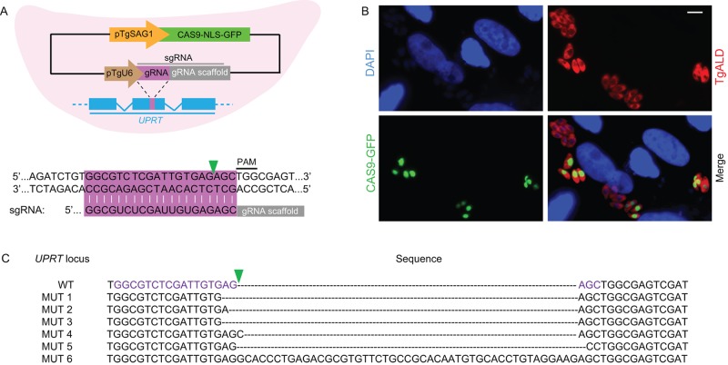
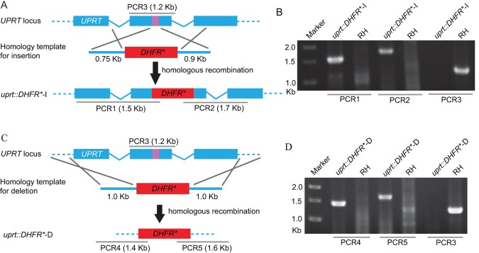
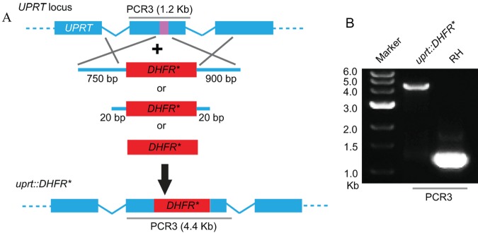
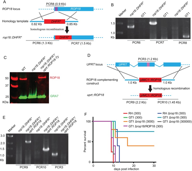
Toxoplasma gondii has become a model for studying the phylum Apicomplexa, in part due to the availability of excellent genetic tools. Although reverse genetic tools are available in a few widely utilized laboratory strains, they rely on special genetic backgrounds that are not easily implemented in natural isolates. Recent progress in modifying CRISPR (clustered regularly interspaced short palindromic repeats), a system of DNA recognition used as a defense mechanism in bacteria and archaea, has led to extremely efficient gene disruption in a variety of organisms. Here we utilized a CRISPR/CAS9-based system with single guide RNAs to disrupt genes in T. gondii. CRISPR/CAS9 provided an extremely efficient system for targeted gene disruption and for site-specific insertion of selectable markers through homologous recombination. CRISPR/CAS9 also facilitated site-specific insertion in the absence of homology, thus increasing the utility of this approach over existing technology. We then tested whether CRISPR/CAS9 would enable efficient transformation of a natural isolate. Using CRISPR/CAS9, we were able to rapidly generate both rop18 knockouts and complemented lines in the type I GT1 strain, which has been used for forward genetic crosses but which remains refractory to reverse genetic approaches. Assessment of their phenotypes in vivo revealed that ROP18 contributed a greater proportion to acute pathogenesis in GT1 than in the laboratory type I RH strain. Thus, CRISPR/CAS9 extends reverse genetic techniques to diverse isolates of T. gondii, allowing exploration of a much wider spectrum of biological diversity.

Importance: Genetic approaches have proven very powerful for studying the biology of organisms, including microbes. However, ease of genetic manipulation varies widely among isolates, with common lab isolates often being the most amenable to such approaches. Unfortunately, such common lab isolates have also been passaged frequently in vitro and have thus lost many of the attributes of wild isolates, often affecting important traits, like virulence. On the other hand, wild isolates are often not amenable to standard genetic approaches, thus limiting inquiry about the genetic basis of biological diversity. Here we imported a new genetic system based on CRISPR/CAS9, which allows high efficiency of targeted gene disruption in natural isolates of T. gondii. This advance promises to bring the power of genetics to bear on the broad diversity of T. gondii strains that have been described recently.


Figures

FIG 1
FIG 1
CRISPR/CAS9-directed mutagenesis in T. gondii. (A) (Top) Schematic illustration of the plasmid expressing CAS9 and a single guide RNA (sgRNA) targeting the UPRT gene in T. gondii. Detailed plasmid map and sequence files can be found in Fig. S1 in the supplemental material. CAS9 (from Streptococcus pyogenes) fused to GFP was expressed from the TgSAG1 promoter, and the sgRNA was expressed from the TgU6 promoter. (Bottom) Sequence of the sgRNA and the region targeted in UPRT (purple bar). The green arrowhead indicates the predicted cleavage site by CAS9. NLS, nuclear localization signal; PAM, protospacer-adjacent motif. (B) Expression of CAS9-NLS-GFP in T. gondii determined by immunofluorescence staining 24 h after transfection of the CAS9/sgUPRT plasmid. Parasites were stained with rabbit anti-TgALD (red), and CAS9-GFP was stained with mouse anti-GFP (green). Scale bar = 5 µm. (C) Mutations in the UPRT gene induced by CAS9 and the sgUPRT from FUDR-resistant clones (MUT 1, etc., represent different clones).
FIG 2
FIG 2
CRISPR/CAS9-mediated gene disruption and/or deletion of the UPRT locus. (A) Schematic of CRISPR/CAS9 strategy used to inactivate UPRT by inserting pyrimethamine-resistant DHFR (DHFR*). Transfection of the sgUPRT together with an amplicon containing a DHFR*-expressing cassette flanked by homology regions to UPRT was used to generate gene disruptions by insertion. The purple bar in UPRT gene represents the region targeted by the sgRNA. (B) Diagnostic PCR demonstrating homologous integration and gene disruption in a representative clone (uprt::DHFR*-I) compared with the parental line RH. PCR1 and PCR2 provide evidence of homologous integration based on products amplified between the DHFR* gene and regions in the UPRT locus that lie outside the targeting amplicon. PCR3 amplified a 1.2-kb fragment in wild-type cells that was lost due to the insertion of DHFR* (the larger fragment created by insertion of DHFR* [4.4 kb] does not amplify under conditions described here). The purple bar in UPRT gene represents the region targeted by the sgRNA. (C) Schematic of CRISPR/CAS9 strategy used to delete the entire coding region of UPRT by inserting DHFR*. Transfection of the sgUPRT together with the DHFR*-expressing amplicon shown was used to generate gene deletions. (D) Diagnostic PCR demonstrating homologous integration and gene deletion in a representative clone (uprt::DHFR*-D) compared with the parental line RH. PCR4 and PCR5 provide evidence of homologous integration based on products amplified between the DHFR* gene and regions in the UPRT locus that lie outside the targeting amplicon. PCR3 was as described above.
FIG 3
FIG 3
Role of flanking regions in CRISPR/CAS9-mediated gene disruption at the UPRT locus. (A) Schematic of CRISPR/CAS9 strategy used to inactivate UPRT by inserting DHFR*. Transfection of the sgUPRT plasmid together with DHFR*-expressing amplicons that contained variable-length flanks with homology to UPRT, or with no homology, were used to generate gene disruptions by insertion. The purple bar in UPRT gene represents the region targeted by the sgRNA. (B) PCR3 analysis of the UPRT locus. Amplification of the endogenous locus (RH) yielded a band of 1.2 kb, while insertion of a single copy of DHFR* resulted in a band of 4.4 kb (uprt::DHFR*). The clone shown was derived by cotransfection of an amplicon of DHFR* with no homology flanks.
FIG 4
FIG 4
CRISPR/CAS9-mediated disruption of ROP18 in the type I strain GT1. (A) Schematic of CRISPR/CAS9-mediated disruption of ROP18 by insertion of DHFR*. Transfection of sgROP18 together with the DHFR* amplicon shown was used to disrupt the ROP18 coding region. The purple bar indicates the sgROP18 target region. (B) Diagnostic PCR demonstrating homologous integration in a representative rop18::DHFR* clone compared with the parental line GT1. (C) Expression of ROP18 in a rop18-disrupted line (rop18::DHFR*) and a complemented line (rop18::DHFR*/uprt::ROP18) determined by Western blotting. ROP18 was detected by rabbit anti-TgROP18, and GRA7 was detected by mouse anti-TgGRA7 as a loading control. Primary antibodies were detected with Li-Cor secondary antibodies and an Odyssey imaging system. (D) Schematic showing the complementation of ROP18-deficient parasites by insertion at the UPRT locus (uprt::ROP18). (E) Diagnostic PCR demonstrating complementation of ROP18 at the UPRT locus (rop18::DHFR*/uprt::ROP18). (F) Survival curves of parasites in CD1 mice. Wild-type RH and GT1 strains were compared to their respective knockouts (∆rop18). The GT1 knockout was complemented to re-express ROP18 (∆rop18/ROP18) at wild-type levels (see panel C). Mice were injected i.p. with the number of parasites indicated in parentheses, and survival was monitored for 30 days.

References

    1. Dubey JP. 2010. Toxoplasmosis of animals and humans. CRC Press, Boca Raton, FL
    1. Sibley LD, Ajioka JW. 2008. Population structure of Toxoplasma gondii: clonal expansion driven by infrequent recombination and selective sweeps. Annu. Rev. Microbiol. 62:329–351. 10.1146/annurev.micro.62.081307.162925 - DOI - PubMed
    1. Hunter CA, Sibley LD. 2012. Modulation of innate immunity by Toxoplasma gondii virulence effectors. Nat. Rev. Microbiol. 10:766–778. 10.1038/nrmicro2858 - DOI - PMC - PubMed
    1. Su C, Khan A, Zhou P, Majumdar D, Ajzenberg D, Dardé ML, Zhu XQ, Ajioka JW, Rosenthal BM, Dubey JP, Sibley LD. 2012. Globally diverse Toxoplasma gondii isolates comprise six major clades originating from a small number of distinct ancestral lineages. Proc. Natl. Acad. Sci. U. S. A. 109:5844–5849. 10.1073/pnas.1203190109 - DOI - PMC - PubMed
    1. Fox BA, Ristuccia JG, Gigley JP, Bzik DJ. 2009. Efficient gene replacements in Toxoplasma gondii strains deficient for nonhomologous end joining. Eukaryot. Cell 8:520–529. 10.1128/EC.00357-08 - DOI - PMC - PubMed

Publication types

Substances