. 2014 Jun 3;53(21):3486-501.
doi: 10.1021/bi500458t. Epub 2014 May 22.

Enzyme architecture: the effect of replacement and deletion mutations of loop 6 on catalysis by triosephosphate isomerase

Affiliations

Enzyme architecture: the effect of replacement and deletion mutations of loop 6 on catalysis by triosephosphate isomerase

Xiang Zhai et al. Biochemistry. .

Abstract

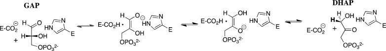
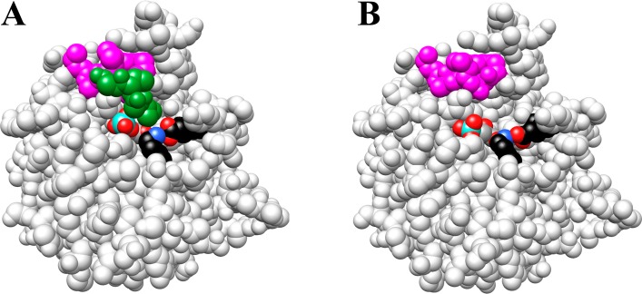
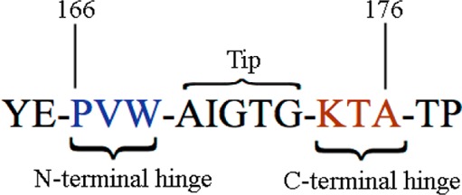
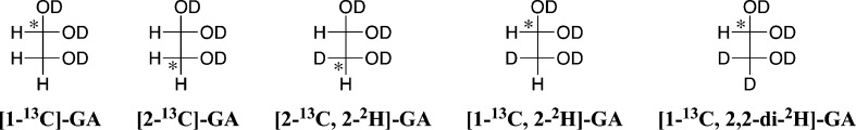
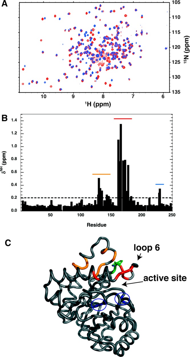
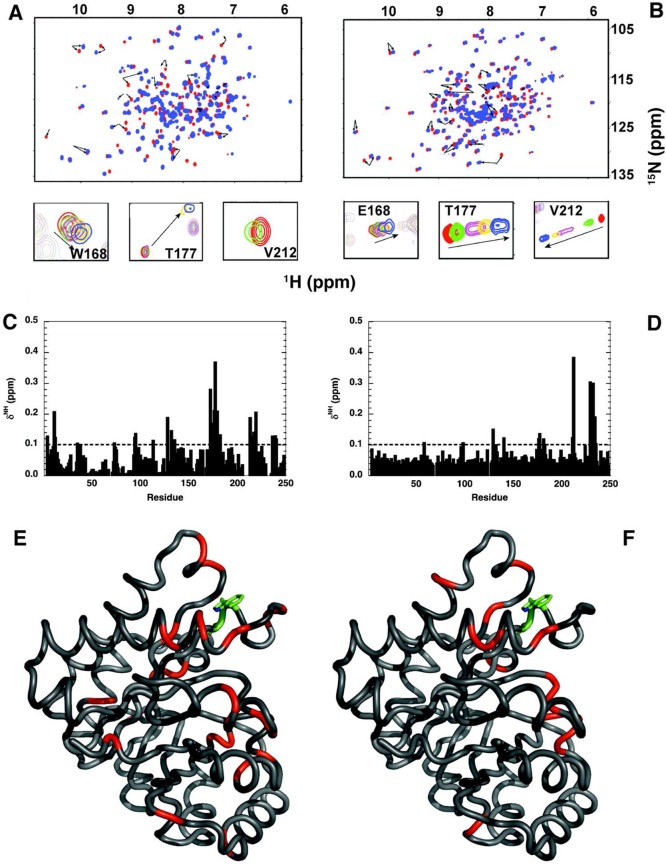
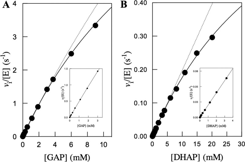
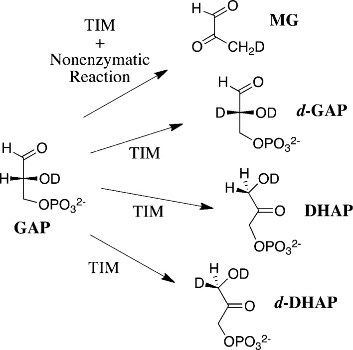
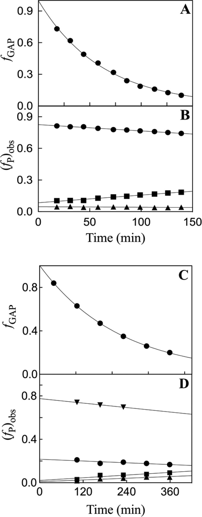
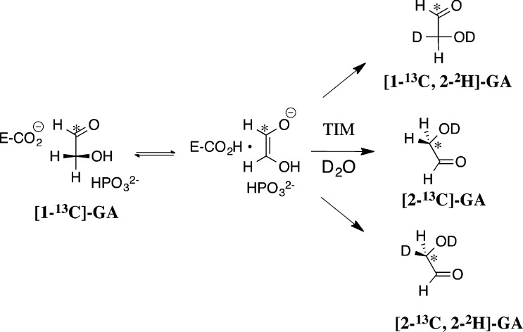
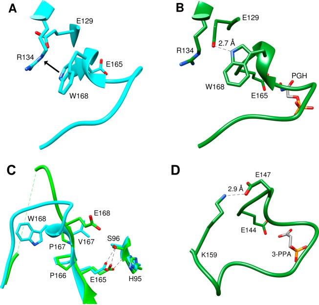
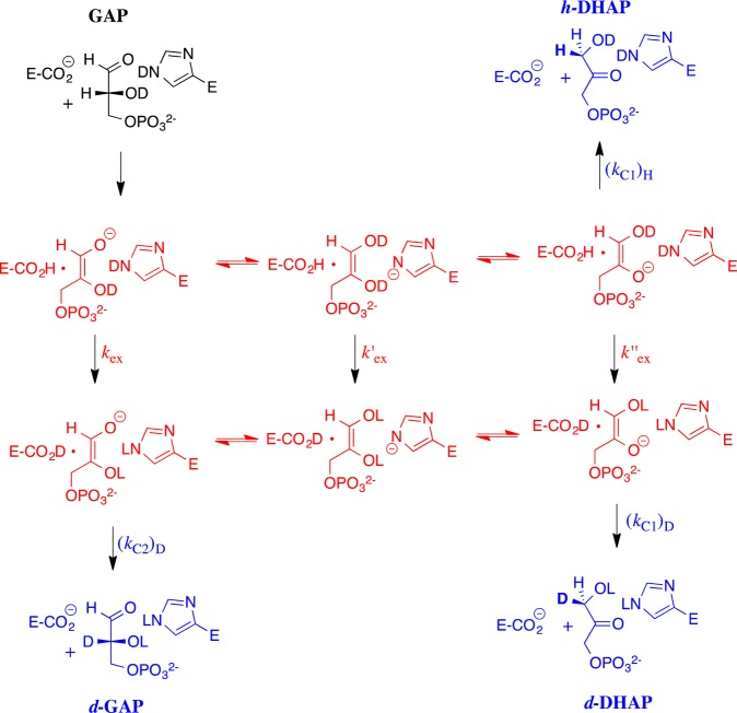
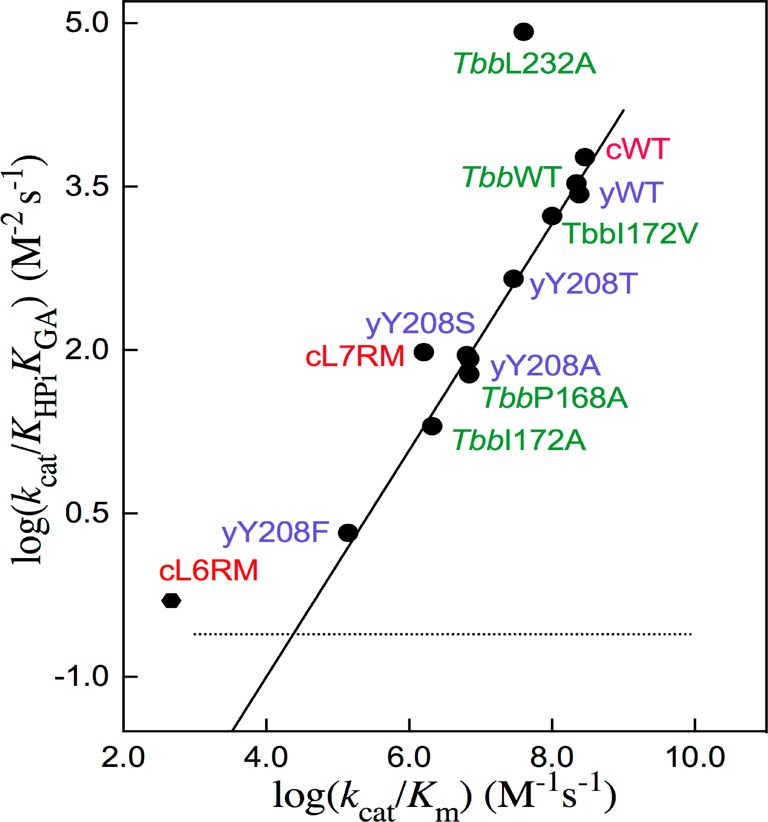
Two mutations of the phosphodianion gripper loop in chicken muscle triosephosphate isomerase (cTIM) were examined: (1) the loop deletion mutant (LDM) formed by removal of residues 170-173 [Pompliano, D. L., et al. (1990) Biochemistry 29, 3186-3194] and (2) the loop 6 replacement mutant (L6RM), in which the N-terminal hinge sequence of TIM from eukaryotes, 166-PXW-168 (X = L or V), is replaced by the sequence from archaea, 166-PPE-168. The X-ray crystal structure of the L6RM shows a large displacement of the side chain of E168 from that for W168 in wild-type cTIM. Solution nuclear magnetic resonance data show that the L6RM results in significant chemical shift changes in loop 6 and surrounding regions, and that the binding of glycerol 3-phosphate (G3P) results in chemical shift changes for nuclei at the active site of the L6RM that are smaller than those of wild-type cTIM. Interactions with loop 6 of the L6RM stabilize the enediolate intermediate toward the elimination reaction catalyzed by the LDM. The LDM and L6RM result in 800000- and 23000-fold decreases, respectively, in kcat/Km for isomerization of GAP. Saturation of the LDM, but not the L6RM, by substrate and inhibitor phosphoglycolate is detected by steady-state kinetic analyses. We propose, on the basis of a comparison of X-ray crystal structures for wild-type TIM and the L6RM, that ligands bind weakly to the L6RM because a large fraction of the ligand binding energy is utilized to overcome destabilizing electrostatic interactions between the side chains of E168 and E129 that are predicted to develop in the loop-closed enzyme. Similar normalized yields of DHAP, d-DHAP, and d-GAP are formed in LDM- and L6RM-catalyzed reactions of GAP in D2O. The smaller normalized 12-13% yield of DHAP and d-DHAP observed for the mutant cTIM-catalyzed reactions compared with the 79% yield of these products for wild-type cTIM suggests that these mutations impair the transfer of a proton from O-2 to O-1 at the initial enediolate phosphate intermediate. No products are detected for the LDM-catalyzed isomerization reactions in D2O of [1-(13)C]GA and HPi, but the L6RM-catalyzed reaction in the presence of 0.020 M dianion gives a 2% yield of the isomerization product [2-(13)C,2-(2)H]GA.


Figures

Scheme 1
Scheme 1
Figure 1
Figure 1
Models that show representations of the X-ray crystal structures of wild-type TIM and a loop 6 deletion mutant (LDM). (A) Space filling model of the complex between TIM from yeast and PGA (PDB entry 2YPI). The amino acid side chains of loop 6 that were retained for the LDM are colored magenta and the deleted residues green. The cationic side chain of K12 is shown with the nitrogen (blue) in an ion pair to oxygen (red) of the anionic side chain of E97. (B) LDM of cTIM generated from the structure of wild-type cTIM by a procedure similar to that described for the K12G mutant of TIM from yeast. The amino acid side chains of loop 6 that were retained for the LDM are colored magenta. Reprinted with permission from ref (17). Copyright 2012 American Chemical Society. In solution, TIM exists as a homodimer. Here we show only the monomer for the sake of clarity.
Scheme 2
Scheme 2
Chart 1
Chart 1
Figure 2
Figure 2
Summary of the effect of the L6RM on the NMR chemical shifts of backbone amide resonances, for spectra acquired at 14.1 T, 298 K, and pH 6.6. (A) Superposition of the resonances for wild-type (red) and L6RM (blue) TIM at the unliganded enzymes. (B) Values of δNH (parts per million) for the L6RM mutation, where δNH is the effect of the L6RM on the composite chemical shift, as defined by eq 3. (C) Model for wild-type cTIM, which shows the positions in the protein structure where the L6RM was found to result in significant values for δNH (>0.2 ppm). The structure has been coded to match the colors shown by the dashed line in panel B with blue residues highlighted by circles. The side chains of the two mutated residues are depicted. The positions of the active site and loop 6 are indicated to orient the viewer.
Figure 3
Figure 3
Summary of the effect of the binding of G3P to wild-type cTIM and to the L6RM on the NMR chemical shifts of backbone amide resonances, for spectra acquired at 14.1 T, 298 K, and pH 6.6. (A and B) Superposition of signals for wild-type cTIM (A) or the L6RM (B) in the unliganded (red) and G3P-saturated (blue) forms. The inset shows the titration profile for residues 168, 177, and 212. The arrows indicate the direction of the shift in the resonance as the unliganded enzyme is saturated by G3P. (C and D) Values of δNH (parts per million) observed upon saturation of wild-type cTIM (C) and the L6RM (D) by G3P, where δNH is the effect of ligand binding on the chemical shift. (E and F) Model for wild-type cTIM (PDB entry 1TIM), which shows, in red, the position in the protein structure where the binding of G3P results in significant values for δNH (>0.1 ppm) for wild-type cTIM (E) or the L6RM (F). The site of the mutation is colored green in panel F.
Figure 4
Figure 4
Michaelis–Menten plots of initial velocity data for the isomerization of GAP and DHAP catalyzed by the L6RM of cTIM at pH 7.5 (30 mM TEA buffer), 25 °C, and I = 0.1 (NaCl). The solid line shows the fit of data to the Michaelis–Menten equation, and the dashed line is the linear relationship of the data at a low substrate concentration (≤3 mM). The inset shows the linear correlation of the initial velocity data for ≤3 mM GAP or DHAP, the slope of which gives the second-order rate constant (kcat/Km).
Scheme 3
Scheme 3
Figure 5
Figure 5
Rate and product data for the reactions of GAP (10 mM) in D2O catalyzed by 0.4 μM L6RM cTIM (A and B) and by 7 μM LDM cTIM (C and D), determined by 1H NMR spectroscopy. (A and C) Decrease in the fraction of GAP (fGAP) remaining for reactions catalyzed by L6RM and LDM cTIM, respectively. (B and D) Fractional product yields, (fP)obs (eq 4), for reactions catalyzed by L6RM and LDM cTIM, respectively. Key for panel B: (●) (fd-DGAP)obs, (■) (fd-DHAP)obs, and (▲) (fDHAP)obs. Key for panel D: (▼) (fMG)obs, (●) (fd-DGAP)obs, (■) (fd-DHAP)obs, and (▲) (fDHAP)obs.
Scheme 4
Scheme 4
Scheme 5
Scheme 5
Figure 6
Figure 6
Representations of X-ray crystal structures of TIM in the region of the N-terminal hinge of loop 6. (A) Wild-type cTIM (PDB entry 1TIM). (B) Wild-type cTIM liganded to PGH (PDB entry 1TPH). (C) Superimposed X-ray crystal structures of unliganded cTIM in the region of the N-terminal hinge: blue ribbon, wild-type cTIM (PDB entry 1TIM); green ribbon, unliganded L6RM of cTIM (PDB entry 4P61). Monomer B, shown for the L6RM, has no significant electron density from residue 173 to 175. (D) TIM from P. woesei liganded to 3-phosphonopropanoic acid (PDB entry 1HG3). The carboxylate side chain of hinge residue E147, which occupies a position equivalent to that of W168 from cTIM, is stabilized by a hydrogen bond to the cationic side chain of K159.
Scheme 6
Scheme 6
Figure 7
Figure 7
Scheme showing the minimal mechanism for the TIM-catalyzed reactions of GAP in D2O that results in the formation of h-DHAP, d-DHAP, and d-GAP. The -H derived from the substrate may exchange with a pool of deuterium at the enzyme. There is evidence that the transfer of hydrogen between O-1 and O-2 of the enediolate reaction intermediate is mediated by the imidazole side chain of His95.
Figure 8
Figure 8
Linear free energy relationship, with a slope of 1.04 ± 0.03, between logarithmic values of second-order rate constants [log(kcat/Km)GAP] for wild-type and mutant TIM-catalyzed isomerization of GAP and the third-order rate constants [log(kcat/KHPiKGA)] for wild-type and mutant TIM-catalyzed reactions of the substrate pieces GA and HPi (Scheme 5). Most of these data were reported and discussed in an earlier publication. The dotted line shows an estimate for the smallest third-order rate constant (kcat/KHPiKGA) that could have been detected by our experimental methods. Key for mutants of TIM: green for TIM from Trypanosoma brucei, red for TIM from chicken muscle, and blue for TIM from yeast.

References

    1. Fothergill-Gilmore L. A. (1986) The evolution of the glycolytic pathway. Trends Biochem. Sci. 11, 47–51.
    1. Knowles J. R.; Albery W. J. (1977) Perfection in enzyme catalysis: The energetics of triosephosphate isomerase. Acc. Chem. Res. 10, 105–111.
    1. Knowles J. R. (1991) To build an enzyme. Philos. Trans. R. Soc. London, Ser. B 332, 115–121. - PubMed
    1. Waley S. G.; Miller J. C.; Rose I. A.; O’Connell E. L. (1970) Identification of site in triose phosphate isomerase labeled by glycidol phosphate. Nature 227, 181. - PubMed
    1. De la Mare S.; Coulson A. F. W.; Knowles J. R.; Priddle J. D.; Offord R. E. (1972) Active-site labeling of triose phosphate isomerase. Reaction of bromohydroxyacetone phosphate with a unique glutamic acid residue and the migration of the label to tyrosine. Biochem. J. 129, 321–331. - PMC - PubMed

Publication types

MeSH terms

LinkOut - more resources