

Early detection of Alzheimer's disease using PiB and FDG PET
- PMID: 24825318
- PMCID: PMC4226742
- DOI: 10.1016/j.nbd.2014.05.001
Early detection of Alzheimer's disease using PiB and FDG PET
Abstract
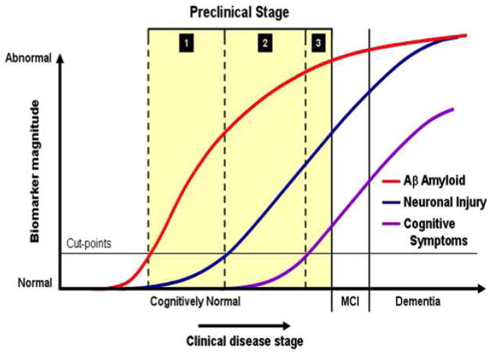
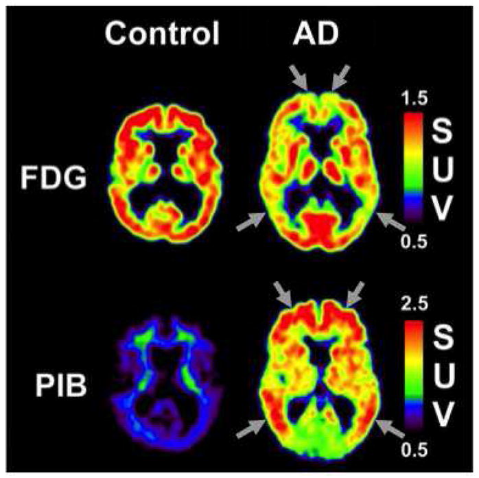
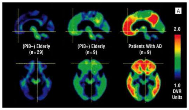
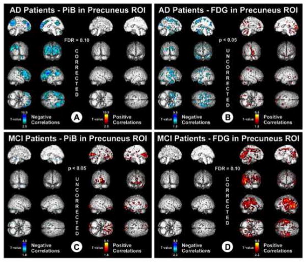
Use of biomarkers in the detection of early and preclinical Alzheimer's disease (AD) has become of central importance following publication of the NIA-Alzheimer's Association revised criteria for the diagnosis of AD, mild cognitive impairment (MCI) and preclinical AD. The use of in vivo amyloid imaging agents, such a Pittsburgh Compound-B and markers of neurodegeneration, such as fluoro-2-deoxy-D-glucose (FDG) is able to detect early AD pathological processes and subsequent neurodegeneration. Imaging with PiB and FDG thus has many potential clinical benefits: early or perhaps preclinical detection of disease and accurately distinguishing AD from dementias of other etiologies in patients presenting with mild or atypical symptoms or confounding comorbidities in which the diagnostic distinction is difficult to make clinically. From a research perspective, this allows us to study relationships between amyloid pathology and changes in cognition, brain structure, and function across the continuum from normal aging to MCI to AD. The present review focuses on use of PiB and FDG-PET and their relationship to one another.
Keywords: Alzheimer's disease; Amyloid; FDG; Glucose metabolism; Neuroimaging; Pittsburgh compound B.
Copyright © 2014 Elsevier Inc. All rights reserved.
Figures
References
-
- Albert MS, et al. The diagnosis of mild cognitive impairment due to Alzheimer’s disease: recommendations from the National Institute on Aging-Alzheimer’s Association workgroups on diagnostic guidelines for Alzheimer’s disease. Alzheimer’s & dementia : the journal of the Alzheimer’s Association. 2011;7:270–9. - PMC - PubMed
-
- Anchisi D, et al. Heterogeneity of brain glucose metabolism in mild cognitive impairment and clinical progression to Alzheimer disease. Arch Neurol. 2005;62:1728–33. - PubMed
-
- Archer HA, et al. Amyloid load and cerebral atrophy in Alzheimer’s disease: an 11C-PIB positron emission tomography study. Ann Neurol. 2006;60:145–7. - PubMed
-
- Arnaiz E, et al. Impaired cerebral glucose metabolism and cognitive functioning predict deterioration in mild cognitive impairment. Neuroreport. 2001;12:851–5. - PubMed
Publication types
MeSH terms
Substances
Grants and funding
LinkOut - more resources
Full Text Sources
Other Literature Sources
Medical
