. 2014 Jul;28(7):1055-72.
doi: 10.1210/me.2013-1134. Epub 2014 May 13.

Androgen-dependent sertoli cell tight junction remodeling is mediated by multiple tight junction components

Affiliations

Androgen-dependent sertoli cell tight junction remodeling is mediated by multiple tight junction components

Papia Chakraborty et al. Mol Endocrinol. 2014 Jul.

Abstract

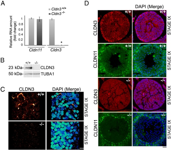
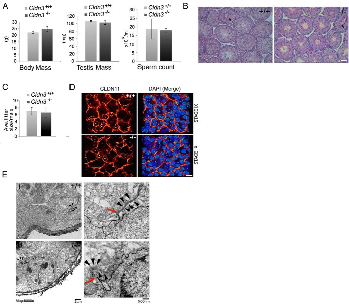
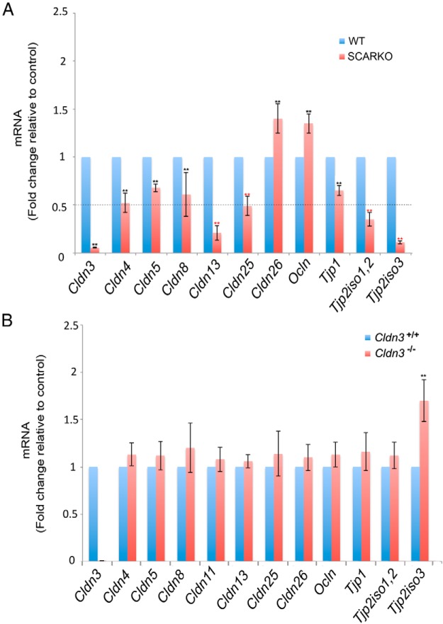
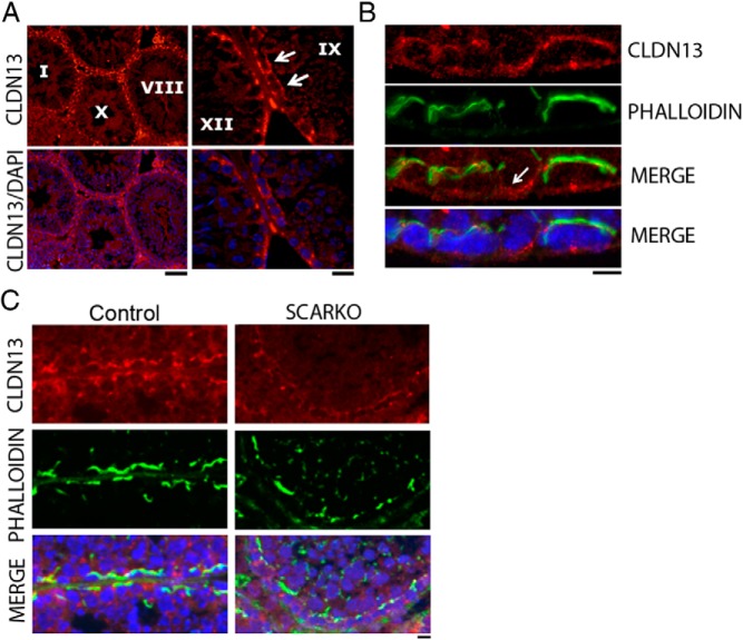
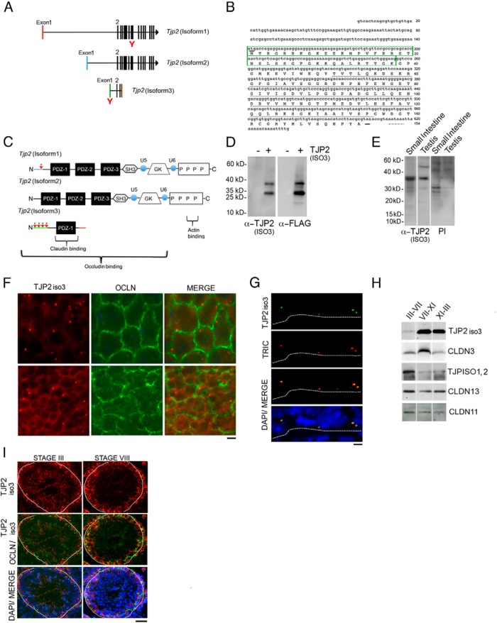
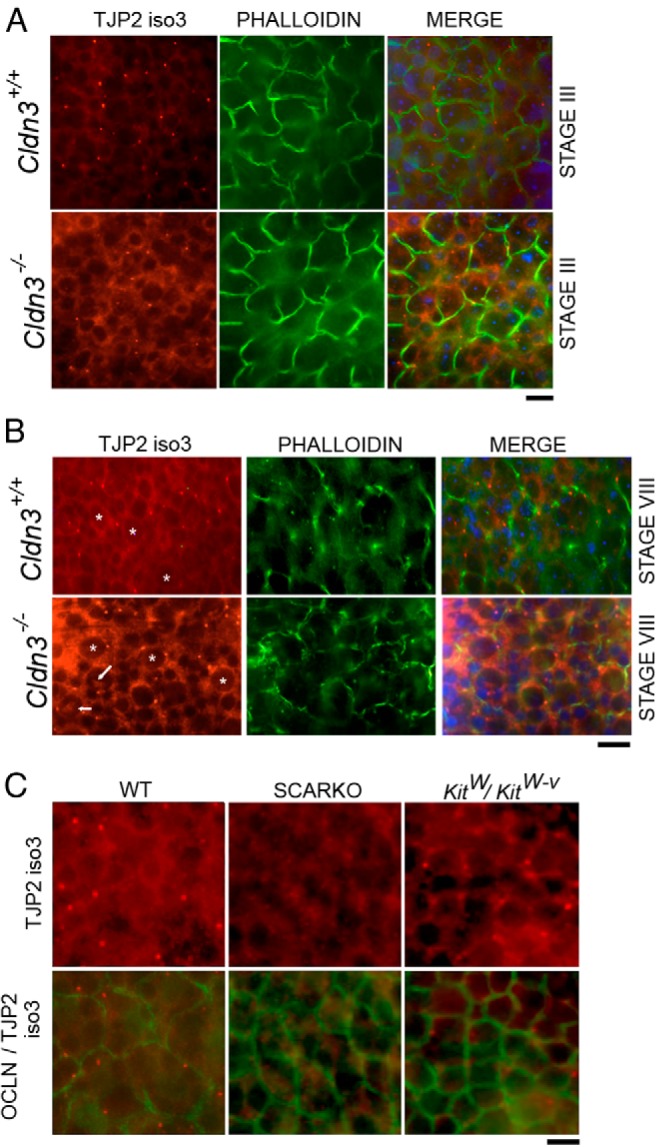
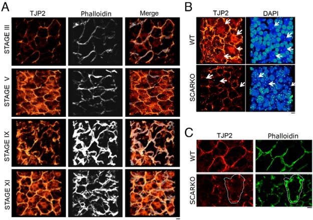
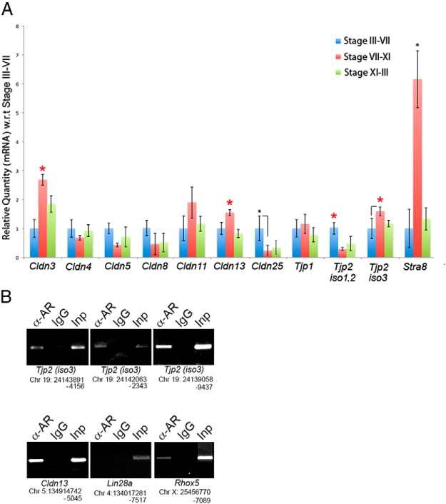
Sertoli cell tight junctions (SCTJs) of the seminiferous epithelium create a specialized microenvironment in the testis to aid differentiation of spermatocytes and spermatids from spermatogonial stem cells. SCTJs must be chronically broken and rebuilt with high fidelity to allow the transmigration of preleptotene spermatocytes from the basal to adluminal epithelial compartment. Impairment of androgen signaling in Sertoli cells perturbs SCTJ remodeling. Claudin (CLDN) 3, a tight junction component under androgen regulation, localizes to newly forming SCTJs and is absent in Sertoli cell androgen receptor knockout (SCARKO) mice. We show here that Cldn3-null mice do not phenocopy SCARKO mice: Cldn3(-/-) mice are fertile, show uninterrupted spermatogenesis, and exhibit fully functional SCTJs based on imaging and small molecule tracer analyses, suggesting that other androgen-regulated genes must contribute to the SCARKO phenotype. To further investigate the SCTJ phenotype observed in SCARKO mutants, we generated a new SCARKO model and extensively analyzed the expression of other tight junction components. In addition to Cldn3, we identified altered expression of several other SCTJ molecules, including down-regulation of Cldn13 and a noncanonical tight junction protein 2 isoform (Tjp2iso3). Chromatin immunoprecipitation was used to demonstrate direct androgen receptor binding to regions of these target genes. Furthermore, we demonstrated that CLDN13 is a constituent of SCTJs and that TJP2iso3 colocalizes with tricellulin, a constituent of tricellular junctions, underscoring the importance of androgen signaling in the regulation of both bicellular and tricellular Sertoli cell tight junctions.


Figures

Figure 1.
Figure 1.
Loss of CLDN3 transcripts and protein in Cldn3−/− testes. A, RT-qPCR analysis of Cldn3 and Cldn11 transcripts from adult testis of Cldn3−/− mice and littermate controls showing a loss of Cldn3 transcript. Error bars represent SEM. n = 3. *, P ≤ .01. B, Western blot analysis of total testis protein extracts prepared from control and Cldn3−/− mice probed with anti-CLDN3 antibody. α-tubulin (TUBA1) antibody was used as a loading control. C, A 3-D confocal reconstruction of a stage IX whole-mount seminiferous tubule immunostained for CLDN3 (red to yellow heat map) and counterstained with DAPI (DNA, blue to green heat map). CLDN3 staining is absent in Cldn3−/− seminiferous tubules. Scale bar corresponds to 10 μm. D, Immunofluorescence detection of CLDN3 (red) and CLDN11 (green) proteins on adult testis sections from control (+/+) and Cldn3−/− mice. CLDN3 protein was absent in Cldn3−/− stage IX tight junctions marked by CLDN11. Staging was determined by DAPI counterstaining (blue). Scale bar corresponds to 10 μm.
Figure 2.
Figure 2.
Qualitative and quantitative spermatogenesis persists in the absence of CLDN3, and SCTJs remain functionally intact. A, Body mass, testis mass, and epididymal sperm counts from 10-week-old control and Cldn3−/− males. n = 5. Error bars represent SEM. No difference between body mass, testis mass, and sperm numbers was detected. B, PAS-stained testis cross sections from 10-week-old control and Cldn3−/− mice. Cldn3−/− testis sections confirm the presence of all cell types and normal spermiation (arrow) in stage VIII tubules. Scale bar corresponds to 30 μm. C, When mated to 12-week-old control females; adult Cldn3−/− males produced litter sizes comparable to those of control males. n = 3. Error bars represent SEM. D, A 3-D confocal reconstruction of a stage IX whole-mount seminiferous tubule immunostained for CLDN11 (red to bright yellow heat map) to identify SCTJs. Nuclei were counterstained with DAPI (blue to green heat map). Migrating preleptotene spermatocytes were observed enclosed within the SCTJs in stage IX (marked by asterisks) of the seminiferous epithelial cycle in both control and Cldn3−/− mice. Scale bar corresponds to 10 μm. E, Electron micrographs of lanthanum colloid tracer studies. Electron microscopy analysis was performed on control and Cldn3−/− testes, after perfusion with lanthanum nitrate (tracer). The lanthanum tracer (black) permeates across the incomplete system of myoid cell layer tight junctions into the intercellular space between Sertoli cells and spermatogonia (insets I and III), where it is blocked by the SCTJs (red arrows). Insets (II and IV) show boxed regions magnified with representative SCTJs (black arrowheads). Diffusion of the tracer across the SCTJ barrier into the adluminal compartment was prevented in control and Cldn3−/− testes. Red arrows mark the point at which the diffusion of the tracer is restricted by the SCTJs.
Figure 3.
Figure 3.
Misregulation of TJP–encoding genes in SCARKO and Cldn3−/− testes. A, Transcript levels of abundant tight junction components were assessed by RT-qPCR analysis from adult control and SCARKO mice. Transcript levels were normalized to Cldn11 and the fold change in mRNA was calculated relative to that of control samples. A >50% reduction in transcripts of Cldn3, Cldn13, Cldn25, Tjp2iso1,2, and Tjp2iso3 was observed in SCARKO testes, whereas Ocln and Cldn26 transcripts were increased. Error bars represent SEM. n = 4. *, P ≤ .05. B, RT-qPCR analysis of abundant tight junction transcripts from adult testes of Cldn3−/− mice and littermate controls show a significant increase in Tjp2iso3 transcript levels, whereas other claudin and tight junction–related transcripts are unaltered. Transcript levels were normalized to actin. Error bars represent SEM. n = 4. *, P ≤ .05. WT, wild-type.
Figure 4.
Figure 4.
CLDN13 localizes to SCTJs and is reduced in AR-deficient Sertoli cells. A, Immunofluorescence detection of CLDN13 (red) and DNA (DAPI; blue) on adult testis cross sections. A typical basal SCTJ-type staining pattern was observed for CLDN13 in all stages. Scale bar corresponds to 20 μm. The right panel confirms presence of CLDN13 in stage IX tubules (white arrow). Scale bar corresponds to 10 μm. B, Immunofluorescence detection of CLDN13 (red), phalloidin (green), and DNA (DAPI; blue) on adult testis cross sections. CLDN13 colocalizes with phalloidin at tight junctions and at subcellular regions not associated with tight junction structures (arrow). Scale bar corresponds to 5 μm. C, Immunofluorescence detection of CLDN13 (red), phalloidin (green), and DNA (DAPI; blue) on adult testis cross sections from control and SCARKO mice. CLDN13 levels were reduced in mutant SCTJs, as identified by phalloidin staining. Scale bar corresponds to 10 μm.
Figure 5.
Figure 5.
TJP2iso3, a truncated isoform of TJP2, is present in the cytosol of germ cells and Sertoli cells and also accumulates at tricellular SCTJs. A, Tjp2 exons are indicated as vertical bars. All 3 isoforms have unique first exons, indicated in red, blue, and green for Tjp2iso1, Tjp2iso2, and Tjp2iso3, respectively. Exons 2, 3, and part of exon 4 are common to all 3 isoforms. The orange bar represents the unique region of exon 4 in Tjp2iso3. B, Nucleic acid sequence and inferred protein sequence derived from Tjp2iso3. The unique amino-terminal sequence contains multiple putative PKA and PKC phosphorylation sites predicted by the NetPhos server (boxed in green). The translational start and stop codons are underlined in bold and the poly(A) signal (AATAAA) is marked with a dashed line. The tight junction binding PDZ-1 domain is underlined in black. C, Comparison of protein domains derived from Tjp2 transcript isoforms. TJP2 proteins link tight junctions to peripheral cytoplasmic proteins. Tight junction association at the cell surface occurs via the PDZ domain, whereas interactions with the actin cytoskeleton are mediated by their proline-rich carboxyl-terminal ends. The PDZ-1 domain alone can associate with the C-terminal cytoplasmic tail of claudins, whereas the PDZ-2 domain binds with TJP1 and connexins. The Src homology-3 and the membrane-associated GUK domains aid in the interactions with the cytoskeletal components such as Ras, Srk kinases, and microtubule-associated proteins. The variable domains termed U (unique) 5 and U6 modulate the specificity of these interactions. The smaller Tjp2iso3 contains the PDZ-1 domain and probably retains its ability to associate with claudins at the tight junctions but lacks domains required for association with cytoskeletal components. D, MDCK cells were stably transfected with the FLAG-TJP2iso3 cDNA or control vector. Whole-cell lysates were subjected to immunoblot analysis with anti-FLAG or anti-TJP2iso3 antibodies. The newly generated TJP2iso3 antibody specifically detected the FLAG-TJP2iso3 protein. E, Analysis of endogenous TJP2iso3 in intestinal and testicular tissues by immunoblot analysis using anti-TJP2iso3 antibody or preimmune serum. F, Longitudinal images of two different regions of a whole seminiferous tubule immunostained for TJP2iso3 (red) and occludin (green). All tubule regions showed a punctate staining, whereas others revealed an additional diffuse cytoplasmic staining pattern. Scale bar corresponds to 5 μm. G, Simultaneous detection of tricellulin (red) and FITC-TJP2iso3 (green). The basal lamina of the tubule is outlined with a white dotted line. Scale bar corresponds to 5 μm. H, Immunoblot analysis of tight junction components in extracts from stage dissected seminiferous tubule fragments. TJP2iso3 and CLDN3 show higher protein levels in stages VII to IX, whereas TJP2iso1,2 levels are greater in stages III to VII. I, Immunofluorescence detection of TJP2iso3 (red) and occludin (green) in 8-week-old adult testis cross sections. DAPI (blue) counterstaining was used to identify stages (III and VIII). Scale bar corresponds to 20 μm.
Figure 6.
Figure 6.
TJP2iso3 protein levels are enhanced in Cldn3−/− mutants and diminished in SCARKO mutants. A, Immunofluorescence detection of TJP2iso3 (red) and phalloidin (green) on whole-mount seminiferous tubules from control and Cldn3−/− mice (DAPI = blue). The diffuse cytosolic TJP2iso3 distribution in Cldn3−/− mutants was enhanced in stage III, whereas the punctate bodies were unaltered. B, Fluorescent images of seminiferous tubules at stage VIII showing TJP2iso3 (red) and phalloidin (green) localization, emphasizing the marked increase in cytoplasmic staining in Cldn3−/− mutant Sertoli cells in comparison with that in controls. Sertoli cells were identified by the bright heterochromatic DAPI foci (asterisks). Nucleolar staining (arrows) of TJP2iso3 was observed in many Sertoli cells in Cldn3−/− mutant mice. The images were obtained at an optical plane (as indicated by a discontinuous tight junction pattern) that clearly displays the Sertoli cell cytoplasm. C, Fluorescence staining of seminiferous tubules with TJP2iso3 (red) and occludin (green) showed a significant reduction in punctate staining of TJP2iso3 in SCARKO mice, whereas the punctate TJP2iso3 staining is still present in KitW/Wv mutant tubules. WT, wild-type.
Figure 7.
Figure 7.
TJP2 levels and spatial localization change during the seminiferous cycle and in SCARKO mice. A, Immunofluorescence detection of TJP2 (red to yellow heat map) and phalloidin (gray) by whole-mount immunostaining and 3-D confocal microscopy show that Sertoli cells have TJP2 in tight junctions throughout the seminiferous cycle (stages III, V, IX, and XI). Stage V Sertoli cells show elevated cytoplasmic TJP2 levels, whereas stage XI Sertoli cells have nuclear localized TJP2. The TJP2 antibody recognizes TJP2iso1 and TJP2iso2 but not TJP2iso3. Scale bar corresponds to 10 μm. B, SCARKO tubules have reduced TJP2 nuclear staining (red to yellow heat map). White arrows in the right panel denote Sertoli cell nuclei, displaying the characteristic heterochromatic pattern. Arrows in the left panel depict the position of the nuclei marked by DAPI (blue to green heat map). Scale bar corresponds to 10 μm. C, Disorganized tight junctions (marked by phalloidin, green) in SCARKO tubules show a modest reduction in TJP2. The white dotted line outlines a tight junction surrounding a single Sertoli cell. Scale bar corresponds to 10 μm. WT, wild-type.
Figure 8.
Figure 8.
Transcript analysis of TJP–encoding genes throughout the seminiferous tubule cycle. A, RT-qPCR analysis of tight junction components performed on total RNA obtained from different seminiferous tubules stages as identified by the transillumination technique. Stra8, a gene expressed at high levels in differentiating spermatocytes during stages VII to XI was used as a positive control to validate the tubule stages isolated using transillumination. Error bars denote SEM. n = 3. *, P ≤ .05. Transcript abundance was plotted relative to stage III to VII for each mRNA. Both TJP2iso3 and Cldn13 transcripts were enriched in stages VII to XI, whereas TJP2iso1 and TJPiso2 were enriched in stages III to VII. B, Testes from 8-week-old male mice were subjected to ChIP with nonimmunized rabbit IgG (negative control) and anti-AR antibodies. The PCR reactions were performed with primer pairs spanning putative AREs in the depicted genomic regions of Tjp2iso3, Cldn13, Lin28a (negative control), and Rhox5 (positive control). (Assembly of mouse genome used for design of primer pairs: December 2011, GRCm38/mm10.)

References

    1. Dym M, Fawcett DW. The blood-testis barrier in the rat and the physiological compartmentation of the seminiferous epithelium. Biol Reprod. 1970;3:308–326. - PubMed
    1. Russell L. Movement of spermatocytes from the basal to the adluminal compartment of the rat testis. Am J Anat. 1977;148:313–328. - PubMed
    1. Mruk DD, Cheng CY. Sertoli-Sertoli and Sertoli-germ cell interactions and their significance in germ cell movement in the seminiferous epithelium during spermatogenesis. Endocr Rev. 2004;25:747–806. - PubMed
    1. Tung KS, Teuscher C, Meng AL. Autoimmunity to spermatozoa and the testis. Immunol Rev. 1981;55:217–255. - PubMed
    1. Smith BE, Braun RE. Germ cell migration across Sertoli cell tight junctions. Science. 2012;338:798–802. - PMC - PubMed

Publication types

MeSH terms