. 2014 Jul;28(7):1039-54.
doi: 10.1210/me.2013-1400. Epub 2014 May 13.

Ovarian dendritic cells act as a double-edged pro-ovulatory and anti-inflammatory sword

Affiliations

Ovarian dendritic cells act as a double-edged pro-ovulatory and anti-inflammatory sword

Adva Cohen-Fredarow et al. Mol Endocrinol. 2014 Jul.

Abstract

Ovulation and inflammation share common attributes, including immune cell invasion into the ovary. The present study aims at deciphering the role of dendritic cells (DCs) in ovulation and corpus luteum formation. Using a CD11c-EYFP transgenic mouse model, ovarian transplantation experiments, and fluorescence-activated cell sorting analyses, we demonstrate that CD11c-positive, F4/80-negative cells, representing DCs, are recruited to the ovary under gonadotropin regulation. By conditional ablation of these cells in CD11c-DTR transgenic mice, we revealed that they are essential for expansion of the cumulus-oocyte complex, release of the ovum from the ovarian follicle, formation of a functional corpus luteum, and enhanced lymphangiogenesis. These experiments were complemented by allogeneic DC transplantation after conditional ablation of CD11c-positive cells that rescued ovulation. The pro-ovulatory effects of these cells were mediated by up-regulation of ovulation-essential genes. Interestingly, we detected a remarkable anti-inflammatory capacity of ovarian DCs, which seemingly serves to restrict the ovulatory-associated inflammation. In addition to discovering the role of DCs in ovulation, this study implies the extended capabilities of these cells, beyond their classic immunologic role, which is relevant also to other biological systems.


Figures

Figure 1.
Figure 1.
DCs reside in the ovary before ovulation and accumulate in the newly formed corpus luteum. A, FACS analysis of ovaries from sexually immature, PMSG-primed C57BL/6 female mice recovered before and at 16, 20, and 24 hours after hCG administration. Ovaries were dissociated into a single-cell suspension, stained with anti-CD11c fluorescent antibodies, and subjected to FACS analysis. Ovaries from nontreated mice (without hormones) served as controls. The number of repetitions, each consisting of 6 to 10 ovaries, is indicated at the bottom of the columns. Error bars represent SEM. *, P < .05. B and C, FACS analysis of ovaries recovered at 48 hours after PMSG administration (B) or at 24 hours after hCG administration to PMSG-primed C57BL/6 mice (C). Ovaries were dissociated, immunostained for CD11c, F4/80, and 7AAD, and subjected to FACS analysis. Only large 7AAD-negative (live) cells are presented. Proportions of gated cells of total large ovarian cells are indicated. n = 4 individual experiments, with 3 to 5 animals each. D, Fluorescence microscopy images of ovaries from CD11c-EYFP transgenic mice at different time intervals after hCG administration demonstrating the dynamic accumulation of CD11c-positive cells (green, CD11c-positive cells; red, blood vessels, BSA-Rox; blue, nuclei, Hoechst). NT, ovaries of nontreated sexually immature mice (without hormones). E, Ex vivo 2-photon microscopy image of ovaries recovered from CD11c-EYFP transgenic mice at 48 hours after PMSG administration, demonstrating a small number of CD11c-positive cells between and around preovulatory follicles (green, CD11c-positive cells; red, blood vessels, Qtracker 655 nontargeted quantum dots; blue, nuclei, Hoechst; scale bar corresponds to 100 μm). F, Ex vivo 2-photon microscopy image of ovaries recovered from PMSG-primed CD11c-EYFP transgenic mice at 24 hours after hCG administration demonstrating a large amount of CD11c-positive cells in a newly formed corpus luteum (green, CD11c-positive cells; red, blood vessels, Qtracker 655 nontargeted quantum dots; blue, nuclei, Hoechst; scale bar corresponds to 100 μm). G, Ex vivo 2-photon microscopy image of ovaries recovered from PMSG-primed CD11c-EYFP transgenic mice at 5 hours after hCG administration demonstrating CD11c-positive cells near and within the wall of blood vessels located in the ovarian interstitium (green, CD11c-positive cells; red, blood vessels, Qtracker 655 nontargeted quantum dots; blue, nuclei, Hoechst; scale bar corresponds to 50 μm).
Figure 2.
Figure 2.
Conditional depletion of CD11c-positive cells blocks hCG-induced ovulation. A, Experimental model: conditional depletion of CD11c-positive cells. CD11c-DTR transgenic mice, in which expression of simian DTR is under the control of the DC promoter sequence CD11c, were used to study the function of DCs in vivo. PMSG-primed CD11c-DTR mice were treated with DTX 24 hours before hCG administration, thus depleting the CD11c-positive cells before the onset of the ovulatory response. B, Ovulated oocytes recovered from CD11c-positive cell–depleted mice compared with those from control mice. Sexually immature CD11c-DTR transgenic mice were treated with either DTX or PBS 24 hours before hCG administration. Ovulated oocytes were recovered from their oviducts 24 hours after hCG administration. Each column presents the mean number of ovulated oocytes per ovary (PBS, n = 16 mice; DTX, n = 22 mice). Error bars represent SEM. *, P < .05. C, Ovulated oocytes recovered after local depletion of CD11c-positive cells. DTX (0, 0.1, 0.5, 1, 5, 10, and 20 ng) was injected unilaterally into the ovarian bursa of sexually immature, PMSG-primed CD11c-DTR female mice at 24 hours before hCG administration (injected ovary), whereas the contralateral ovary remained untreated (noninjected ovary). Ovulated oocytes were recovered separately from each oviduct at 16 hours after hCG administration. The effect of DTX administration was dose dependent (P < .05). n = ≥6 mice/group; error bars represent SEM. D, Hematoxylin and eosin–stained histological section of an ovary of a CD11c-depleted mouse showing a trapped oocyte (indicated by black arrow) that resides within the corpus luteum (scale bar corresponds to 100 μm).
Figure 3.
Figure 3.
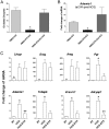
The presence of CD11c-positive DCs in the ovary is essential for ovulation and for the expression of hCG-induced ovulatory genes. A, Allogeneic transplantation of DCs into the ovarian bursa of hCG-stimulated, DTX-treated CD11c-DTR mice restores ovulation. The graph presents the number of oocytes ovulated after hormonal treatment (PMSG-hCG), hormonal treatment after depletion of CD11c-positive cells (PMSG-DTX-hCG), and the combination of hormonal treatment and DTX administration with allogeneic transplantation of DCs into the ovarian bursa (PMSG-DTX-DCs-hCG). Sexually immature CD11c-DTR transgenic mice were treated as described, and ovulated oocytes were recovered from their oviducts 24 hours after hCG administration. Each column presents the mean number of ovulated oocytes per oviduct. n = 15 mice. Error bars represent SEM. *, P < .05. B, Allogeneic transplantation of DCs into the ovarian bursa of hCG-stimulated, DTX-treated CD11c-DTR mice restored Adamts1 mRNA expression. Graph presents quantitative real-time PCR analyses of Adamts1 at 24 hours after hCG administration. Ovaries were recovered from CD11c-DTR transgenic mice after either hormonal treatment (PMSG-hCG), hormonal treatment after the depletion of CD11c-positive cells (PMSG-DTX-hCG), or the combination of hormonal treatment and DTX administration with allogeneic transplantation of DCs into the ovarian bursa (PMSG-DTX-DCs-hCG). n = 15 mice. Error bars represent SEM. *, P < .05. C, Quantitative real-time PCR analyses of genes essential for ovulation at 4 hours after hCG administration. Ovaries were recovered from DC-depleted (hCG-DTX), nondepleted (hCG), and untreated (NT) CD11c-DTR mice. n = ≥4 individual experiments, each including 4 mice/group. Error bars represent SEM. *, P < .05. Columns with different markings (* vs ** vs no marking) are significantly different.
Figure 4.
Figure 4.
Cumulus cell mucification/expansion is depended on the present of CD11c-positive DCs in the ovary. A, Conditional depletion of CD11c-positive cells impaired cumulus cell mucification/expansion. The graph presents quantification of the morphological status of COCs (compact, expended, and denuded; shown in the right panels) as was evaluated by light microscopy. COCs were recovered from either ovaries of PMSG-primed mice with or without hCG administration (at 8 hours) or PMSG-primed mice treated by DTX followed by hCG administration. Most COCs in PMSG-primed mice were compact and expended after hCG administration; however, many of the COCs recovered from CD11c-depleted mice contained denuded oocytes (scale bar corresponds to 100 μm). B and C, Scanning electron microscopy (airSEM; backscattered with beam energy of 30 kV and probe current of 500 pA) of COCs recovered from PMSG-primed CD11c-DTR transgenic, either DTX-treated (C) or DTX-untreated (B), mice at 8 hours after hCG administration. Note the abnormal appearance and low amount of extracellular matrix between the cumulus cells of CD11c-depleted mice (scale bar corresponds to 100 μm (upper images) and 10 μm (bottom images). D, Mass spectrometry protein analysis of VCAN-V1 and TNFAIP6 in the extracellular matrix of COCs recovered from PMSG-primed CD11c-DTR transgenic, either DTX-treated or DTX-untreated, mice at 8 hours after hCG administration.
Figure 5.
Figure 5.
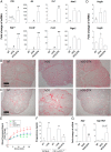
Depletion of CD11c-positive cells alters ovarian inflammatory gene expression, lymphatic vessel development, and progesterone production. A, Quantitative real-time PCR analyses of inflammation-associated genes. Ovaries were recovered at 4 hours after hCG administration from DC-depleted (hCG-DTX), nondepleted (hCG), and untreated (NT) CD11c-DTR mice. n = 4 individual experiments, each including 4 mice/group. Error bars represent SEM. *, P < .05. B, Immunostaining for the blood vessel marker α-SMA performed on ovaries recovered 20 hours after hCG administration from DC-depleted (hCG-DTX), nondepleted (hCG), and untreated (NT) CD11c-DTR mice, demonstrating an hCG-stimulated increase in ovarian blood vessels, particularly in the newly developed corpora lutea, that was not altered by depletion of CD11c-positive cells (n = 5 mice, 10 ovaries/group; scale bar corresponds to 100 μm). C, Immunostaining for LYVE-1 shows a significant increase in the ovarian lymphatic vessels after hCG administration that was severely impaired by the depletion of CD11c-positive cells (n = 5 mice, 10 ovaries/group; scale bar corresponds to 200 μm). D, Quantitative real-time PCR analyses of Vegfa, and Vegfc. Ovaries were recovered at 4 hours after hCG administration from DC-depleted (hCG-DTX), nondepleted (hCG), and untreated (NT) CD11c-DTR mice. n = 4 individual experiments, each including 4 mice/group. Error bars represent SEM. *, P < .05. E, In vivo ovarian fluorescent signal dynamics of BSA-Rox injected intravenously at 24 hours after hCG administration to DC-depleted (hCG-DTX), nondepleted (hCG), and untreated (NT) PMSG-primed CD11c-DTR mice (n = 5 mice/group). For each animal, the ovarian fluorescent signal in each time point was normalized to the initial signal (ie, at 1 minute, associated with blood vessel density). Over time, the signal intensity dynamics in the imaged ovaries of the CD11c-depleted mice demonstrated higher incremental increased rate, indicating higher accumulation of the BSA-Rox in their ovaries. F, Serum progesterone concentrations of either CD11c-DTR (DTR) or wild-type (C57BL/6) mice. Serum progesterone concentrations were evaluated in untreated mice (NT) at 24 hours after hCG administration to either PMSG-primed nondepleted (hCG) or PMSG-primed, CD11c-depleted mice (hCG-DTX). n = 5 mice/group; error bars represent SEM. *, P < .05. G, Quantitative real-time PCR analyses of Star and Cyp19a1. Ovaries were recovered at 4 hours after hCG administration from DC-depleted (hCG-DTX), nondepleted (hCG), and untreated (NT) CD11c-DTR mice. n = 4 individual experiments, each including 4 mice/group. Error bars represent SEM. *, P < .05. Columns with different markings (* vs ** vs no marking) are significantly different.
Figure 6.
Figure 6.
Model depicting the role of DCs in the ovary during ovulation. Upon gonadotropin stimulation, the relatively small ovarian DC population is essential for the LH-stimulated up-regulation of specific ovulatory genes that are crucial for cumulus mucification/expansion and ovulation. As the ovulatory process progresses, the ovarian DC population expands and localizes to the newly formed corpus luteum. During this process, up-regulation of the inflammatory repressing genes confine the inflammatory conditions provoked by ovulation. In the early luteal phase, the substantial amount of DCs localized in the newly formed corpus luteum facilitates progesterone production as well as ovarian lymphangiogenesis.

References

    1. Espey LL. Ovulation as an inflammatory reaction—a hypothesis. Biol Reprod. 1980;22:73–106. - PubMed
    1. Richards JS, Liu Z, Shimada M. Immune-like mechanisms in ovulation. Trends Endocrinol Metab. 2008;19:191–196. - PubMed
    1. Tsafriri A, Lindner HR, Zor U, Lamprecht SA. Physiological role of prostaglandins in the induction of ovulation. Prostaglandins. 1972;2:1–10. - PubMed
    1. Reich R, Kohen F, Slager R, Tsafriri A. Ovarian lipoxygenase activity and its regulation by gonadotropin in the rat. Prostaglandins. 1985;30:581–590. - PubMed
    1. Reich R, Daphna-Iken D, Chun SY, et al. . Preovulatory changes in ovarian expression of collagenases and tissue metalloproteinase inhibitor messenger ribonucleic acid: role of eicosanoids. Endocrinology. 1991;129:1869–1875. - PubMed

Publication types

MeSH terms