. 2014 Sep:74:139-50.
doi: 10.1016/j.yjmcc.2014.05.001. Epub 2014 May 10.

Cardiomyocytes mediate anti-angiogenesis in type 2 diabetic rats through the exosomal transfer of miR-320 into endothelial cells

Affiliations

Cardiomyocytes mediate anti-angiogenesis in type 2 diabetic rats through the exosomal transfer of miR-320 into endothelial cells

Xiaohong Wang et al. J Mol Cell Cardiol. 2014 Sep.

Abstract

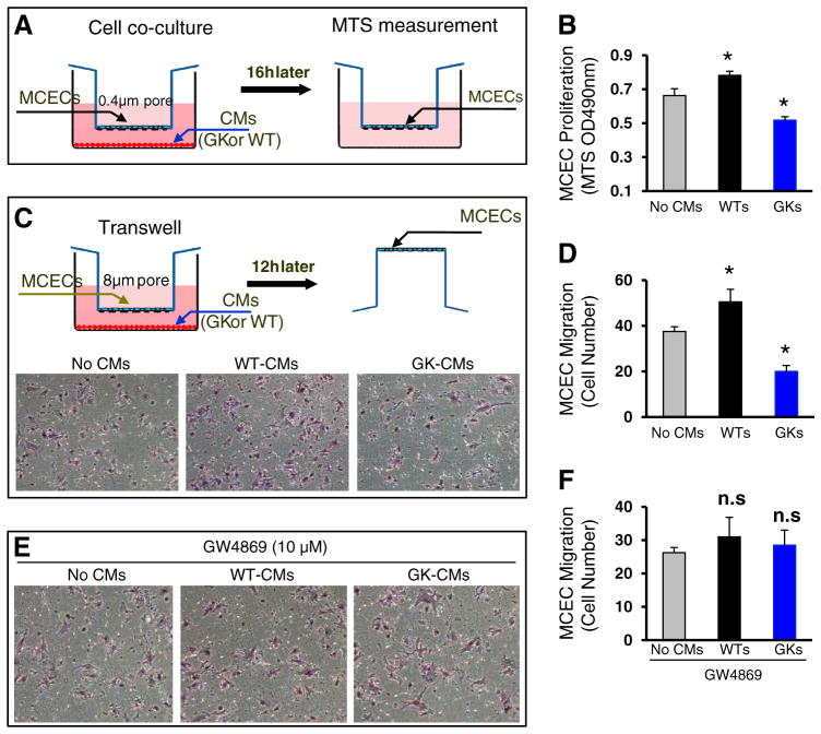
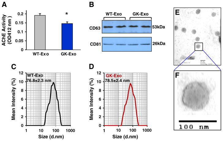
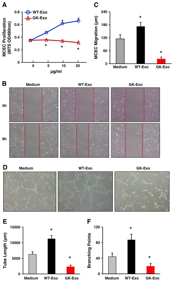
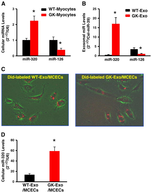
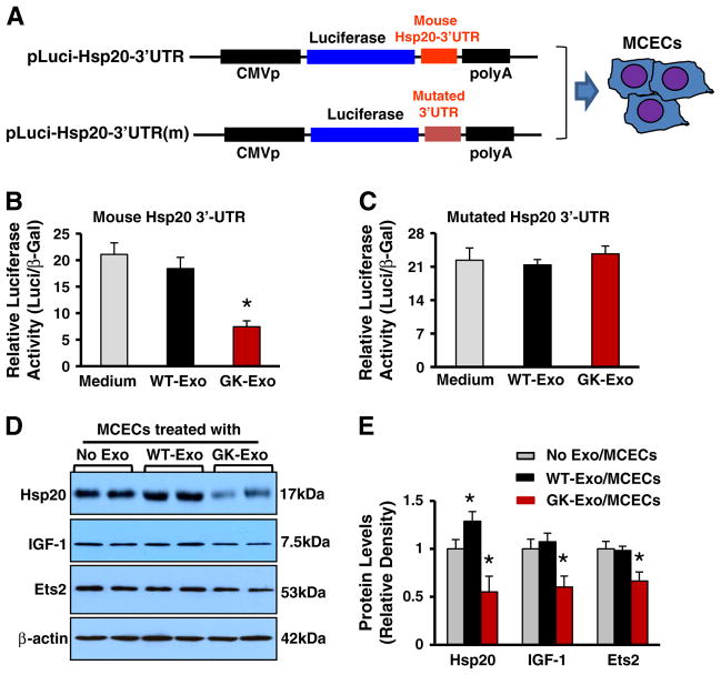
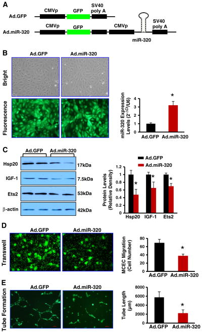
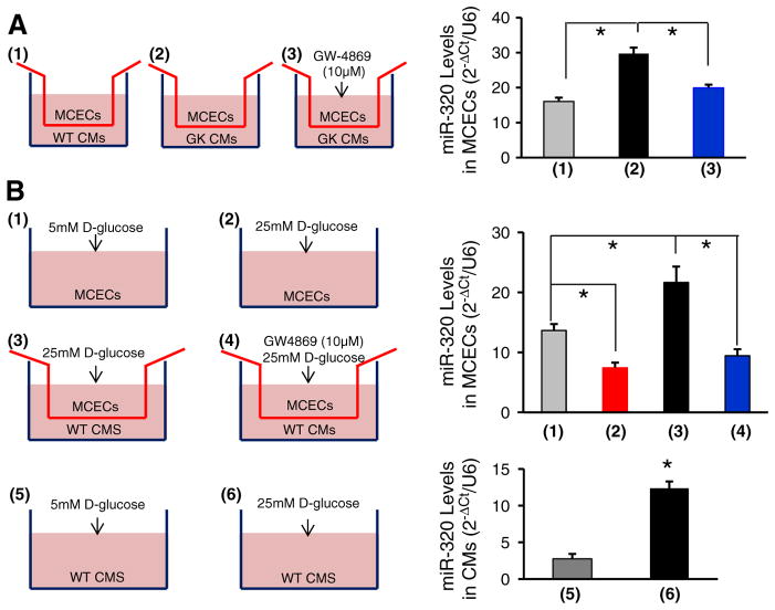
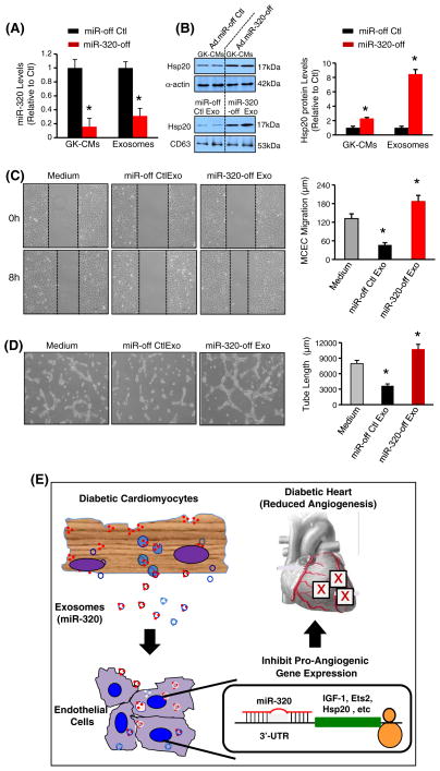
Exosomes, nano-vesicles naturally released from living cells, have been well recognized to play critical roles in mediating cell-to-cell communication. Given that diabetic hearts exhibit insufficient angiogenesis, it is significant to test whether diabetic cardiomyocyte-derived exosomes possess any capacity in regulating angiogenesis. In this study, we first observed that both proliferation and migration of mouse cardiac endothelial cells (MCECs) were inhibited when co-cultured with cardiomyocytes isolated from adult Goto-Kakizaki (GK) rats, a commonly used animal model of type 2 diabetes. However, GK-myocyte-mediated anti-angiogenic effects were negated upon addition of GW4869, an inhibitor of exosome formation/release, into the co-cultures. Next, exosomes were purified from the myocyte culture supernatants by differential centrifugation. While exosomes derived from GK myocytes (GK-exosomes) displayed similar size and molecular markers (CD63 and CD81) to those originated from the control Wistar rat myocytes (WT-exosomes), their regulatory role in angiogenesis is opposite. We observed that the MCEC proliferation, migration and tube-like formation were inhibited by GK-exosomes, but were promoted by WT-exosomes. Mechanistically, we found that GK-exosomes encapsulated higher levels of miR-320 and lower levels of miR-126 compared to WT-exosomes. Furthermore, GK-exosomes were effectively taken up by MCECs and delivered miR-320. In addition, transportation of miR-320 from myocytes to MCECs could be blocked by GW4869. Importantly, the exosomal miR-320 functionally down-regulated its target genes (IGF-1, Hsp20 and Ets2) in recipient MCECs, and overexpression of miR-320 inhibited MCEC migration and tube formation. GK exosome-mediated inhibitory effects on angiogenesis were removed by knockdown of miR-320. Together, these data indicate that cardiomyocytes exert an anti-angiogenic function in type 2 diabetic rats through exosomal transfer of miR-320 into endothelial cells. Thus, our study provides a novel mechanism underlying diabetes mellitus-induced myocardial vascular deficiency which may be caused by secretion of anti-angiogenic exosomes from cardiomyocyes.

Keywords: Cardiomyocytes; Exosomes; Myocardial angiogenesis; Type 2 diabetes; miR-320.


Conflict of interest statement

Conflict of interest disclosures

None.

Figures

Fig. 1
Fig. 1
Proliferation and migration of mouse cardiac endothelial cells (MCECs) are inhibited when co-cultured with cardiomyocytes isolated from GK rats, whereas they are promoted when co-cultured with Wistar (WT) rat cardiomyocytes. (A) A scheme of cell co-culture system in which cardiomyocytes were cultured in the lower chamber of a 12-well plate pre-coated with mouse laminin (10 μg/ml) and MCECs were cultured in the upper chamber of a 12-well insert. (B) GK myocytes inhibited MCEC proliferation, which was promoted by WT myocytes. (C) A scheme of the transwell experiment to evaluate endothelial cell migration. Representative endothelial cells which were trans-welled when co-cultured with WT-cardiomyocytes or GK cardiomyocytes. The quantitative results of endothelial cells migrated are shown in (D). (E/F) Cardiomyocyte-mediated regulatory effects on EC migration were negated upon addition of GW4869. n = 4 wells for each group, and similar results were observed in three additional, independent experiments; *p < 0.05 vs. no cardiomyocyes (CMs).
Fig. 2
Fig. 2
Characterization of exosomes released from WT- and GK-cardiomyocytes. (A) The activity of acetycholinesterase (AChE) was reduced in GK-exosomes collected from the same amount of cardiomyocytes, compared with controls (n = 4 rats, *p < 0.05). (B) The levels of CD63 and CD81 were similar between GK-exosomes and WT-exosomes (100 μg of exosomal protein was loaded for Western-blotting). (C/D) The exosome size was measured using a Zetasizer Nano instrument (n = 4 independent experiments). (E/F) WT-exosomes were examined under electron microscopy.
Fig. 3
Fig. 3
Effects of GK-exosomes on angiogenesis. (A) GK-exosomes significantly inhibited MCEC proliferation in a dose-dependent manner (n = 6 wells, *p < 0.05 vs. WT-exosomes, similar results were observed in three independent experiments). (B/C) GK-exosomes (20 μg/ml) attenuated MCEC migration and (D–F) tube-like formation. Conversely, WT-exosomes(20 μg/ml) exhibited pro-angiogenic effects (B–F). Total capillary tube length (E) and tube branch points (F) were measured by analytical software Image-Pro Plus 5.1. Five independent fields were assessed for each well (n = 3 wells for each group). All values were expressed as means ± SD; *p < 0.05 vs. medium controls. Similar results were observed in three additional, independent experiments.
Fig. 4
Fig. 4
Cardiomyocyte-exosomes uptake and deliver miR-320 in endothelial cells. (A) The levels of miR-320 and miR-126 were examined in (A) cardiomyocytes and (B) myocyte-derived exosomes. (C) Red fluorescent dye DiD-labeled WT-exosomes and GK-exosomes were uptaken and distributed in the cytoplasm of MCECs. (D) The miR-320 levels were dramatically increased in MCECs upon exposure to GK-exosomes. U6 RNA was used as an internal control for qRT-PCR of cellular RNA analysis. C. elegans miR-39 was used as spike-in control for qRT-PCR of exosomal RNA analysis. (n = 5, *p < 0.01 vs. WT-exosomes.)
Fig. 5
Fig. 5
Cardiomyocyte exosomal miR-320 functionally down-regulates its target genes in recipient endothelial cells. (A) Diagrams of plasmid construction in which mouse Hsp20 3′-UTR or its mutation was inserted downstream of the luciferase cDNA. (B/C) Dual luciferase activity assay in MCECs treated with WT-exosomes or GK exosomes. (D/E) Western-blotting assay revealed that the protein levels of Hsp20, IGF-1, and Ets2 were significantly reduced in GK-exosome-treated MCECs. n = 4 independent experiments; *p < 0.05 vs. exosome-null-treated MCECs.
Fig. 6
Fig. 6
Overexpression of miR-320 inhibits MCEC migration and tube formation. (A) Diagrams of recombinant adenoviral vectors (Ad.GFP and Ad.miR-320). (B) MCECs were infected with Ad.GFP or Ad.miR-320, and the miR-320 levels were measured with qRT-PCR. U6 RNA was used as an internal control. (C) The protein levels of Hsp20, IGF-1 and Ets2 were significantly reduced in Ad.miR-320-cells, compared with Ad.GFP-cells. Overexpression of miR-320 greatly inhibited MCEC migration (D) and tube formation (E). *p < 0.05 vs. Ad.GFP, n = 3 independent experiments.
Fig. 7
Fig. 7
Transportation of miR-320 from cardiomyocytes into MCECs is blocked by the exosome inhibitor GW4869. (A) Schemes of MCECs co-cultured with: (1) WT-myocytes, (2) GK-myocytes and (3) GW4869-treated GK-myocytes. Twenty-four hours later, MCECs were collected for the determination of miR-320 levels by qRT-PCR. Addition of GW4869 prevented the increase of miR-320 in MCECs co-cultured with GK-myocytes (*p < 0.05 vs. controls as indicated). Similar results were observed in three additional, independent experiments. (B) Schemes of MCECs: (1) cultured in the presence of 5 mM D-glucose, (2) in the presence of 25 mM D-glucose, (3) co-cultured with WT-cardiomyocytes in the presence of 25 mM D-glucose and (4) co-cultured with WT-cardiomyocytes in the presence of 25 mM D-glucose plus GW4869; schemes of WT cardiomyocytes cultured in the presence of 5 mM D-glucose (5) and in the presence of 25 mM D-glucose (6). The levels of miR-320 were reduced in MCECs under high glucose conditions (2), whereas its levels were remarkably increased when co-cultured with high-glucose-treated cardiomyocytes (3). Such an increase of miR-320 levels may have originated from cardiomyocytes, as miR-320 levels were increased in WT cardiomyocytes upon high-glucose treatment (6), and addition of the exosome inhibitor GW4869 blocked the transfer of miR-320 from cardiomyocytes into MCECs (4). (*p < 0.05 vs. controls as indicated). Similar results were observed in three additional, independent experiments.
Fig. 8
Fig. 8
Effects of miR-320-reduced exosomes collected form miR-320-off GK cardiomyocytes on angiogenesis. (A) Infection of GK-cardiomyocytes with Ad.miR-320-off dramatically reduced miR-320 levels in cardiomyocytes and their released exosomes. (B) Protein levels of Hsp20 were remarkably increased in Ad.miR-320-off-infected GK myocytes and their released exosomes. n = 4, *p < 0.05 vs. controls indicated. miR-320-off-exosomes (20 μg/ml) significantly promoted (C) MCEC migration and (D) tube-like formation, compared with miR-off control exosomes. Total capillary tube length was measured as described in Fig. 3. All values were expressed as means ± SD; *p < 0.05 vs. medium controls. Similar results were observed in three additional, independent experiments. (E) Proposed scheme for cardiomyocyte-mediated anti-angiogenesis through exosomal miR-320 in diabetic hearts.

Comment in

References

    1. Raslova K. An update on the treatment of type 1 and type 2 diabetes mellitus: focus on insulin detemir, a long-acting human insulin analog. Vasc Health Risk Manag. 2010;6:399–410. - PMC - PubMed
    1. Nakagami H, Kaneda Y, Ogihara T, Morishita R. Endothelial dysfunction in hyperglycemia as a trigger of atherosclerosis. Curr Diabetes Rev. 2005;1:59–63. - PubMed
    1. Cohen G, Riahi Y, Alpert E, Gruzman A, Sasson S. The roles of hyperglycaemia and oxidative stress in the rise and collapse of the natural protective mechanism against vascular endothelial cell dysfunction in diabetes. Arch Physiol Biochem. 2007;113:259–67. - PubMed
    1. Costa PZ, Soares R. Neovascularization in diabetes and its complications. Unraveling the angiogenic paradox. Life Sci. 2013;92:1037–45. - PubMed
    1. Heather LC, Clarke K. Metabolism, hypoxia and the diabetic heart. J Mol Cell Cardiol. 2011;50:598–605. - PubMed

Publication types

MeSH terms