. 2014 Jul 24;124(4):611-22.
doi: 10.1182/blood-2014-02-554980. Epub 2014 May 13.

Platelets are required for enhanced activation of the endothelium and fibrinogen in a mouse thrombosis model of APS

Affiliations

Platelets are required for enhanced activation of the endothelium and fibrinogen in a mouse thrombosis model of APS

Valerie Proulle et al. Blood. .

Abstract

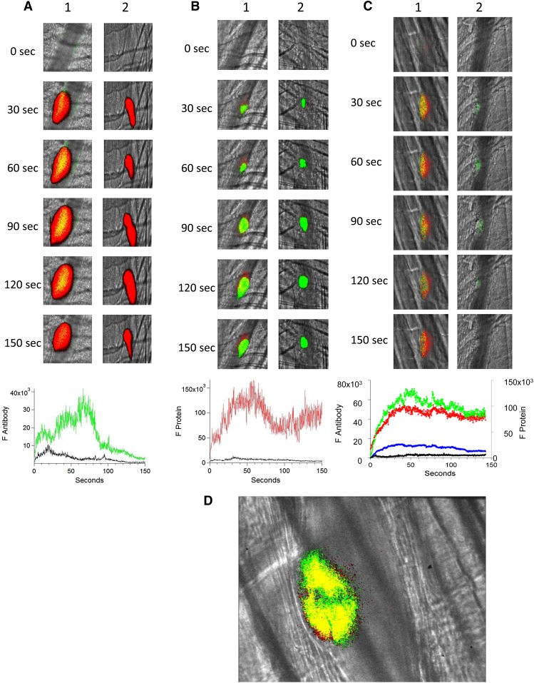
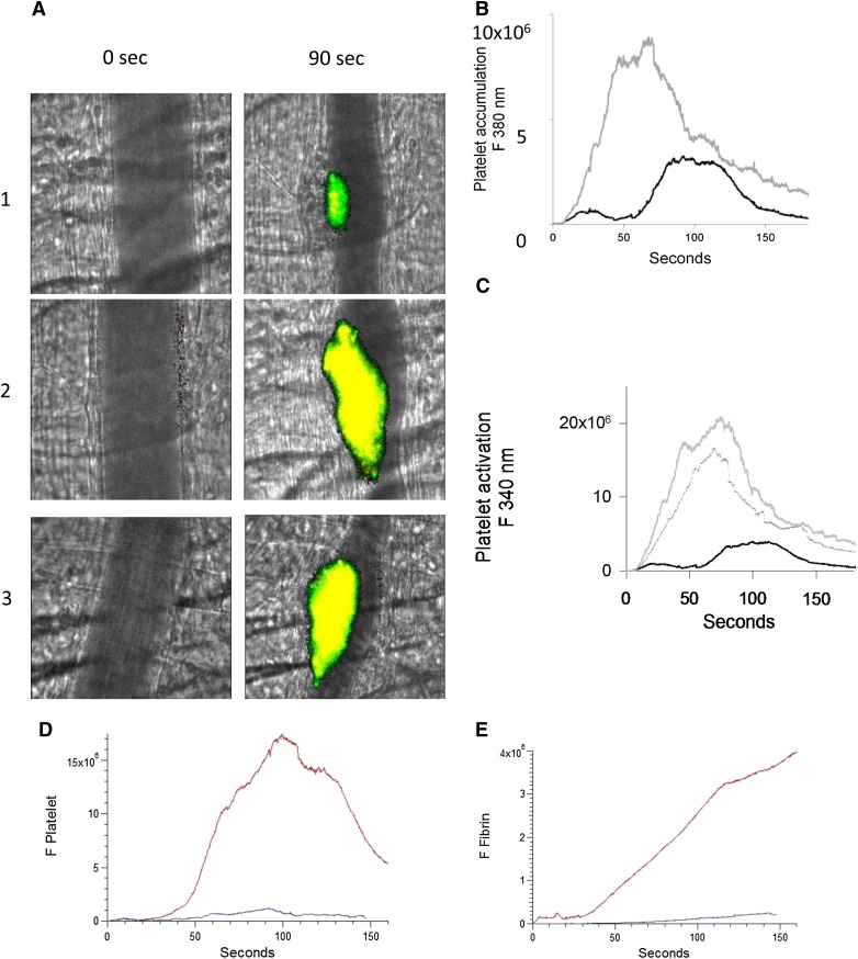
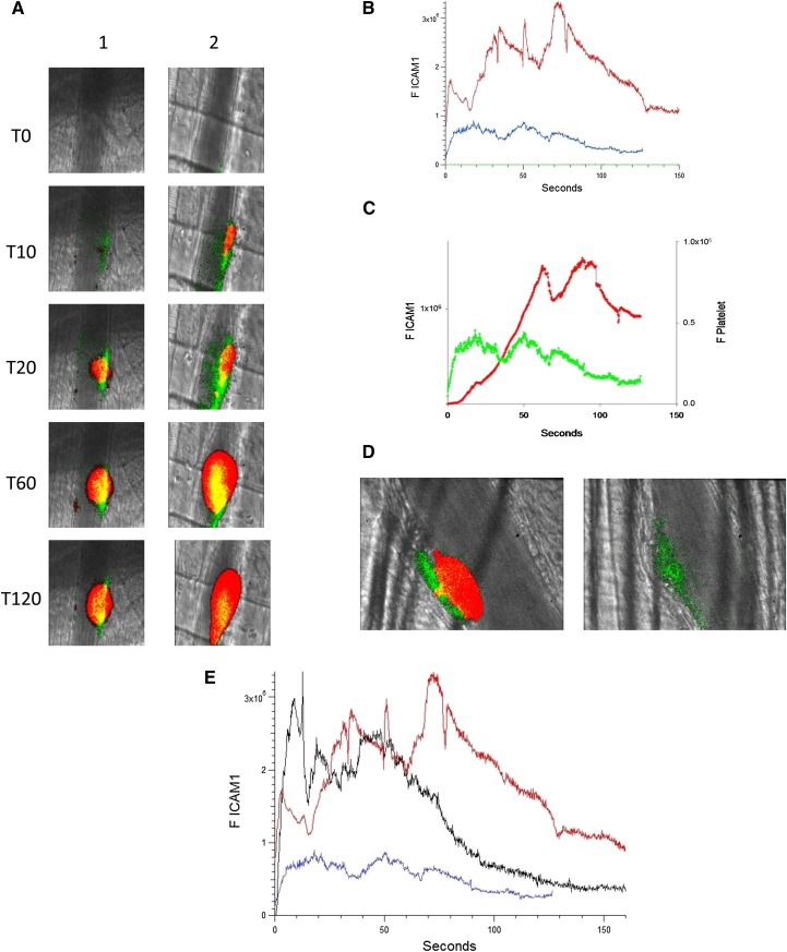
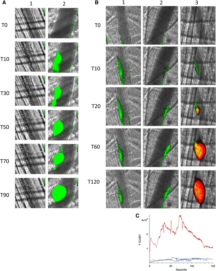
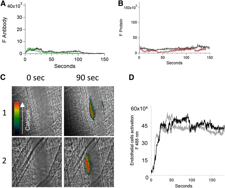
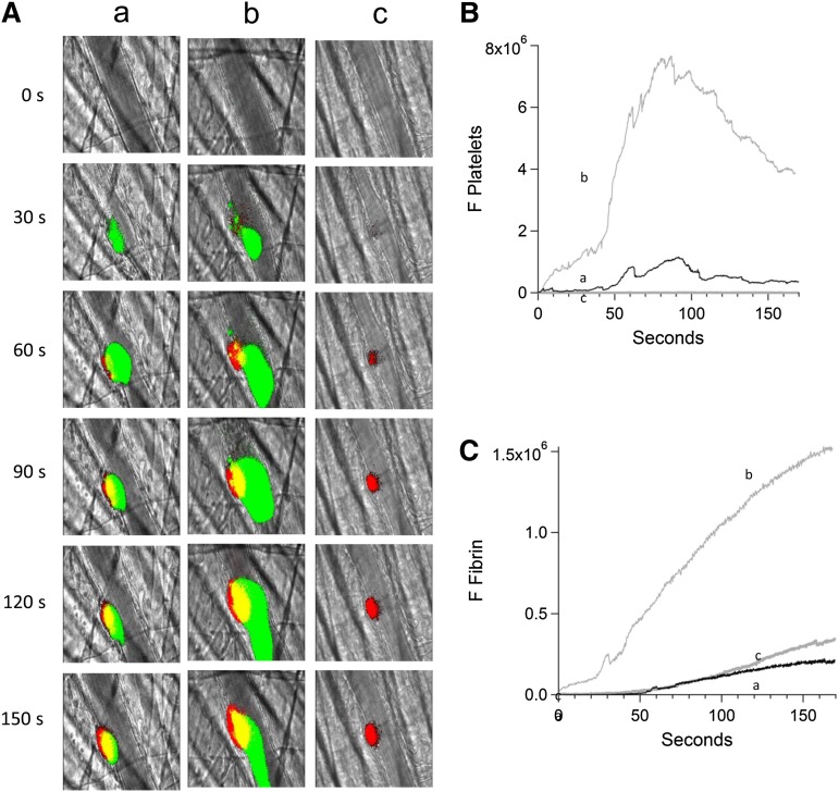
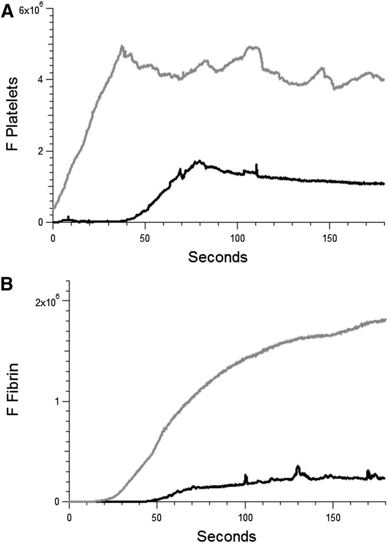
Antiphospholipid syndrome (APS) is defined by thrombosis, fetal loss, and the presence of antiphospholipid antibodies, including anti-β2-glycoprotein-1 autoantibodies (anti-β2GP1) that have a direct role in the pathogenesis of thrombosis in vivo. The cellular targets of the anti-β2GP1 autoantibody/β2GP1 complex in vivo were studied using a laser-induced thrombosis model of APS in a live mouse and human anti-β2GP1 autoantibodies affinity-purified from APS patients. Cell binding of fluorescently labeled β2GP1 and anti-β2GP1 autoantibodies revealed their colocalization on the platelet thrombus but not the endothelium. Anti-β2GP1 autoantibodies enhanced platelet activation, monitored by calcium mobilization, and endothelial activation, monitored by intercellular adhesion molecule-1 expression. When eptifibatide was infused to block platelet thrombus formation, enhanced fibrin generation and endothelial cell activation were eliminated. Thus, the anti-β2GP1 autoantibody/β2GP1 complex binds to the thrombus, enhancing platelet activation, and platelet secretion leads to enhanced endothelium activation and fibrin generation. These results lead to a paradigm shift away from the concept that binding of the anti-β2GP1 autoantibody/β2GP1 complex activates both endothelial cells and platelets toward one in which activation of platelets in response to anti-β2GP1 autoantibody/β2GP1 complex binding leads to subsequent enhanced endothelium activation and fibrin generation.


Figures

Figure 1
Figure 1
Binding of fluorescently labeled human anti-β2GP1 F(ab′)2, fluorescently labeled β2GP1, or both to the developing thrombus. (A) Binding of fluorescently labeled human anti-β2GP1 F(ab′)2 to the developing thrombus. Anti-β2GP1 F(ab′)2 (2.5 μg/mouse) or control F(ab′)2 (2.5 μg/mouse), each labeled with Alexa 488, and anti-CD42 antibody labeled with Dylight 649 were infused into the mouse 15 minutes before laser-induced arteriolar wall injury. The results shown are for binding of anti-β2GP1 F(ab′)2 derived from sera from APS patient 1. Similar results were obtained with anti-β2GP1 F(ab′)2 derived from sera from APS patient 2. (Top) Representative images of the fluorescence signal associated with anti-β2GP1 F(ab′)2 (green; lane 1) or control F(ab′)2 (green; lane 2) and platelets (red) over 150 seconds after vessel injury are shown within the context of the brightfield histology. Merge (yellow). (Bottom) The median integrated antibody fluorescence (FANTIBODY) associated with thrombus formation after infusion of anti-β2GP1 F(ab′)2 (51 thrombi, 5 mice) or control F(ab′)2 (51 thrombi, 4 mice) over 150 seconds after vessel wall injury. Anti-β2GP1 F(ab′)2 (green); control F(ab′)2 (black). (B) Binding of fluorescently labeled β2GP1 in the presence of human anti-β2GP1 autoantibodies to the developing thrombus. Mice were infused with β2GP1 (25 μg/mouse) or HSA (25 μg/mouse), each labeled with Alexa 647, anti-β2GP1 autoantibodies (10 μg/mouse), and anti-CD42 antibody labeled with Dylight 488 before laser-induced arteriolar wall injury. (Top) Representative images of the fluorescence signal associated with β2GP1 (lane 1, red) or HSA (lane 2, red) and anti-CD42 antibody labeled with Dylight 488 for platelet detection (green) over 150 seconds after vessel injury are shown within the context of the brightfield histology. Merge (yellow). (Bottom) Median integrated protein fluorescence (Fβ2GP1 or FHSA) associated with thrombus formation in 3 wild-type mice after infusion of β2GP1 conjugated to Alexa 647 (27 thrombi, 3 mice) or HSA control conjugated to Alexa 647 (24 thrombi, 3 mice) over 150 seconds after vessel wall injury. β2GP1 (red); HSA (black). (C) Simultaneous binding of fluorescently labeled anti-β2GP1 autoantibodies and fluorescently labeled β2GP1 during thrombus formation after laser-induced injury. Mice were infused with labeled anti-β2GP1 F(ab′)2 (2.5 μg) derived from patient 2 plus labeled β2GP1 (25 μg) or labeled control F(ab′)2 (2.5 μg) plus labeled HSA (25 μg) 15 minutes before laser-induced arteriolar wall injury. (Top) Representative images of the fluorescence signal associated with Alexa 488-labeled anti-β2GP1 F(ab′)2 (lane 1, green) or Alexa 488–labeled control F(ab′)2 (lane 2, green) and Alexa 647–labeled β2GP1 (lane 1, red) or Alexa 647–labeled HSA (lane 2, red) over 150 seconds after vessel injury are shown within the context of the brightfield histology. Merge (yellow). (Bottom) The median integrated fluorescence, (FANTIBODY) and (FPROTEIN), of antibody and protein, respectively, associated with thrombus formation after infusion of Alexa 488–labeled anti-β2GP1 F(ab′)2 and Alexa 647–labeled β2GP1 (25 thrombi, 3 mice) or Alexa 488–labeled control F(ab′)2 and Alexa 647–labeled HSA (26 thrombi, 2 mice) over 150 seconds after vessel wall injury. Anti-β2GP1 F(ab′)2 (green); control F(ab′)2 (blue); β2GP1 (red); HSA (black). (D) High-resolution confocal intravital imaging of binding of the anti-β2GP1 autoantibody/β2GP1 complex during thrombus formation after laser-induced injury. Confocal image of thrombus formation 60 seconds after vessel wall injury. Alexa 488–labeled anti-β2GP1 F(ab′)2 (green); β2GP1 (red); merge (yellow). A single confocal slice through the center of the thrombus is shown.
Figure 2
Figure 2
Enhanced platelet activation at the site of vascular injury with infusion of anti-β2GP1 autoantibodies. Mice were infused with platelets loaded with fura-2-AM (250 × 106/mouse). Monitoring fluorescence at 380 nm allowed quantitation of platelet accumulation (unliganded fura-2, green) and monitoring fluorescence at 340 nm allowed quantitation of activated platelets (Ca+2 liganded fura-2, red; yellow = merge). (A) Images of the developing thrombus at time 0 and at 90 seconds obtained without (panel 1), with infusion of 10 μg anti-β2GP1 IgG (panel 2) and with infusion of 12 μg anti-β2GP1 F(ab′)2 (panel 3). Resting platelets, green; activated platelets, yellow. (B) Platelet accumulation after laser-induced injury is represented by the median fluorescence intensity of loaded platelets excited at 380 nm over 3 minutes in 20 thrombi in 4 mice before (black) and in 25 thrombi after (gray) infusion of 10 μg of anti-β2GP1 IgG. (C) Platelet activation at the site of laser-induced injury is represented by the median fluorescence intensity of fura-2-AM–loaded platelets excited at 340 nm over 3 minutes in 20 thrombi in 4 mice before (black) and 25 thrombi after (gray) infusion of 10 μg of anti-β2GP1 IgG. (D) F(ab′)2 of anti-β2GP1 IgG-induced enhancement of platelet accumulation. (E) F(ab′)2 of anti-β2GP1 IgG-induced enhancement of fibrin generation. Platelet accumulation and fibrin generation at the site of laser-induced injury were measured before and 20 minutes after infusion of 12 μg of F(ab′)2 of anti-β2GP1 IgG. Platelet and fibrin labeling were performed using anti-CD42 antibody labeled with Dylight 488 (0.1 μg/g) and anti-fibrin antibody labeled with Alexa 647 (0.5 μg/g). F(ab′)2 of anti-β2GP1 IgG-induced changes in platelet thrombus size and fibrin generation were observed in thrombi performed upstream in a single arteriole before (blue, 16 thrombi, 3 mice) and 20 minutes after infusion of 12 μg of F(ab′)2 of anti-β2GP1 IgG (red, 12 thrombi, 2 mice).
Figure 3
Figure 3
Endothelial cell activation is enhanced at the site of vascular injury in the presence of anti-β2GP1 autoantibodies in vivo. (A) Representative images of ICAM-1 and platelets in the developing thrombus from time 0 to 120 seconds obtained before (left panel, lane 1) and 15 minutes after (right panel, lane 2) infusion of 10 μg of human anti-β2GP1 autoantibodies. ICAM-1, green; platelets, red; merge, yellow. (B) The median integrated ICAM-1 fluorescence (F ICAM-1) associated with thrombus formation before (19 thrombi, 4 mice; blue) and 15 minutes after (27 thrombi, 4 mice; red) infusion of 10 μg of anti-β2GP1 IgG over 150 seconds after vessel wall injury. An irrelevant IgG in place of the anti-ICAM-1 antibody is shown (21 thrombin, 2 mice; green). (C) Comparison of the kinetics of ICAM-1 expression (green) and platelet accumulation (red) during thrombus formation in the absence of anti-β2GP1 IgG. (D) Confocal imaging of ICAM-1 after vascular injury indicates ICAM-1 is localized on the endothelium and not the platelet thrombus. Confocal images of ICAM-1 and platelets were obtained 60 seconds after laser injury during thrombus formation. (Left) ICAM-1 was visualized using anti-ICAM-1 labeled with Alexa 488 (0.4 μg/g mouse) (green) and platelets were visualized using anti-CD42 antibody labeled with Dylight 649 (0.1 μg/g mouse) (red). Merge, yellow. (Right) In the presence of eptifibatide (10 μg/g mouse) and its elimination of platelets, ICAM-1 was visualized on the endothelial surface. Confocal images were obtained through a central section of the thrombus. These confocal images are obtained at high speed in a live mouse where there is minor vessel movement with during each systole. Furthermore, the green and the red images are obtained near simultaneously but not simultaneously. Therefore, the register of the composite image is not perfect. Finally, there is low background noise that we elected not to subtract. We quantitated the fluorescence corresponding to total ICAM-1 fluorescence in the image and quantitated the fluorescence corresponding to the ICAM-1 fluorescence within the thrombus, as defined by platelet fluorescence. (E) F(ab′)2 fragments of anti-β2GP1 autoantibodies enhance activation of endothelial cells similarly to intact anti-β2GP1 autoantibodies. Endothelial cell activation was monitored using anti-ICAM-1 conjugated to Alexa 488 (0.5 μg/g) and platelets were monitored using anti-CD42 antibody conjugated with Dylight 649 (0.1 μg/g) before and 20 minutes after infusion of 12 μg of F(ab′)2 fragments of anti-β2GP1. ICAM-1 expression in endothelial cells was observed in an arteriole before (blue; 19 thrombi, 4 mice) and 20 minutes after infusion of 12 μg of F(ab′)2 fragments of anti-β2GP1 (black; 14 thrombi, 2 mice) or 10 μg of intact anti-β2GP1 (red; 27 thrombi, 4 mice).
Figure 4
Figure 4
Platelet thrombus is required for enhanced endothelial cell activation in the presence of anti-β2GP1 autoantibodies. (A) Infusion of eptifibatide (10 μg/g mouse) initially inhibits platelet interaction with the injured vessel wall but its effect is gone by 60 minutes. Platelets were labeled using anti-CD42 antibody conjugated to Dylight 488 (0.1 μg/g) and platelet fluorescence (green) imaged at 488 nm. Images were obtained with the high-resolution CMOS camera that resolves individual platelets. (Lane 1) Image at time 0. (Lane 2) Image at time 60 minutes after eptifibatide infusion. (B) Representative images of ICAM-1 and platelets in the developing thrombus from time 0 to 120 seconds obtained in the presence of eptifibatide (10 μg/g mouse) (lane 1); human anti-β2GP1 autoantibodies (10 μg) and eptifibatide (10 μg/g mouse) (lane 2); human anti-β2GP1 autoantibodies (10 μg) in the absence of eptifibatide (lane 3). ICAM-1, green; platelets, red. (C) The median integrated ICAM-1 fluorescence (F ICAM-1) associated with thrombus formation in wild-type mice infused with human anti-β2GP1 autoantibodies (27 thrombi, 4 mice; red); human anti-β2GP1 autoantibodies and eptifibatide (27 thrombi, 3 mice; blue); and eptifibatide (18 thrombi, 3 mice; black).
Figure 5
Figure 5
In the absence of a platelet thrombus, the anti-β2GP1 autoantibody/β2GP1 complex neither bound the endothelium nor enhanced endothelial calcium mobilization. (A) After blocking platelet accumulation by infusion of eptifibatide (10 μg/g mouse), the median integrated antibody fluorescence (FAntibody) after infusion of anti-β2GP1 F(ab′)2 (24 thrombi, 2 mice) or control F(ab′)2 (26 thrombi, 2 mice) over 150 seconds after vessel wall injury was measured. Anti-β2GP1 F(ab′)2 (green); control F(ab′)2 (black). No significant binding of anti-β2GP1 F(ab′)2 was observed in the absence of a platelet thrombus. (B) After blocking platelet accumulation by infusion of eptifibatide (10 μg/g mouse), the median integrated protein fluorescence (Fβ2GP1 or FHSA) after infusion of Alexa 647 conjugated to β2GP1 (24 thrombi, 2 mice) or Alexa 647 conjugated to HSA (26 thrombi, 2 mice) over 150 seconds after vessel wall injury was measured. β2GP1 (red); control (black). No significant binding of β2GP1 was observed. (C) Fluo-4-AM was delivered into the mouse circulation via the femoral artery, and concurrent platelet aggregation was inhibited by infusion of eptifibatide (10 μg/g mouse) every 15 minutes. After laser-induced vessel wall injury, changes to endothelial Ca2+ levels were observed by excitation at 488 nm and images were recorded over time. Antibody-induced change in endothelial cell activation was examined in the injured vessel performed upstream in 1 arteriole before and 15 minutes after infusion of 10 μg of anti-β2GP1 autoantibodies. Images show calcium elevation in the endothelium in the absence of platelet accumulation obtained at time 0 and at 90 seconds after vessel wall injury before (panel 1) and after (panel 2) injection of 10 μg of anti-β2GP1 antibodies. The fluorescence signal is represented as a pseudocolor intensity map where black represents the least intense and red represents the most intense fluorescence signal. (D) The kinetics of endothelial cell activation at the site of laser-induced injury was determined by calculating median fluorescence values at 488 nm as a function of time. Calcium mobilization is represented by the median fluorescence intensity of Fluo-4-AM–loaded endothelial cells over 3 minutes before (black) and after (gray) infusion of 10 μg of anti-β2GP1 IgG for 15 to 20 thrombi in 2 mice.
Figure 6
Figure 6
Inhibition of platelet thrombus formation with eptifibatide prevents anti-β2GP1 autoantibody–mediated enhancement of fibrin generation. Platelet and fibrin imaging were performed using anti-CD42 antibody labeled with Dylight 488 (0.1 μg/g mouse) and anti-fibrin antibody labeled with Alexa 647 (0.5 μg/g mouse). Platelet thrombus size and fibrin generation at the site of laser-induced injury were determined by calculating median fluorescence values at 488 nm and 647 nm over 3 minutes, respectively. Anti-β2GP1 autoantibody–induced changes in platelet thrombus size and fibrin generation were observed in thrombi performed upstream in a single arteriole before (16 thrombi, 3 mice) and 15 minutes after (20 thrombi, 3 mice) infusion of 10 μg of anti-β2GP1 antibodies. Subsequently, platelet accumulation at the site of injury was prevented by infusion of eptifibatide (10 μg/g mouse) every 15 minutes, and platelet thrombus size and fibrin generation were observed in thrombi (19 thrombi, 3 mice) performed upstream in a single arteriole in the presence of anti-β2GP1 autoantibodies. (A) Representative images of the developing thrombus obtained (a) without antibody, (b) with 10 μg anti-β2GP1 IgG, and (c) with 10 μg anti-β2GP1 IgG and eptifibatide. Platelets, green; fibrin, red; merge, yellow. (B) Platelet accumulation. (C) Fibrin generation. No antibody (a), black; anti-β2GP1 autoantibody (b), light gray; anti-β2GP1 autoantibody and eptifibatide (c), dark gray.
Figure 7
Figure 7
Anti-β2GP1 autoantibodies amplify thrombus formation in venules. Effect of purified anti-β2GP1 autoantibodies on thrombus size and fibrin generation in venules. Anti-β2GP1 autoantibodies were infused into wild-type mice 5 minutes before laser-induced venule wall injury. Platelet and fibrin imaging was performed using anti-CD42 antibody labeled with Dylight 649 (0.1 μg/g mouse) and anti-fibrin antibody labeled with Alexa 488 (0.5 μg/g mouse). Platelet thrombus size and fibrin generation at the site of laser-induced injury were determined by calculating median fluorescence values at 649 nm and 488 nm over 3 minutes, respectively. After initial laser injury of the venule wall, a thrombus composed of platelets (A) and fibrin (B) was generated; 15 minutes after infusion of 10 μg of anti-β2GP1 autoantibodies, anti-β2GP1 autoantibody–induced changes in platelet thrombus size and fibrin generation were observed. The kinetics of the fluorescence signals associated with platelets and fibrin over 180 seconds after vessel injury are shown before (black) and after (gray) infusion of anti-β2GP1 autoantibodies.

Comment in

References

    1. de Groot PG, Urbanus RT. The significance of autoantibodies against β2-glycoprotein I. Blood. 2012;120(2):266–274. - PubMed
    1. Willis R, Harris EN, Pierangeli SS. Pathogenesis of the antiphospholipid syndrome. Semin Thromb Hemost. 2012;38(4):305–321. - PubMed
    1. Meroni PL, Borghi MO, Raschi E, Tedesco F. Pathogenesis of antiphospholipid syndrome: understanding the antibodies. Nat Rev Rheumatol. 2011;7(6):330–339. - PubMed
    1. McNeil HP, Simpson RJ, Chesterman CN, Krilis SA. Anti-phospholipid antibodies are directed against a complex antigen that includes a lipid-binding inhibitor of coagulation: beta 2-glycoprotein I (apolipoprotein H). Proc Natl Acad Sci U S A. 1990;87(11):4120–4124. - PMC - PubMed
    1. Iverson GM, Victoria EJ, Marquis DM. Anti-beta2 glycoprotein I (beta2GPI) autoantibodies recognize an epitope on the first domain of beta2GPI. Proc Natl Acad Sci U S A. 1998;95(26):15542–15546. - PMC - PubMed

Publication types

MeSH terms