. 2014 Aug 28;124(9):1460-72.
doi: 10.1182/blood-2014-03-559542. Epub 2014 May 13.

Integrated genomic sequencing reveals mutational landscape of T-cell prolymphocytic leukemia

Affiliations

Integrated genomic sequencing reveals mutational landscape of T-cell prolymphocytic leukemia

Mark J Kiel et al. Blood. .

Abstract

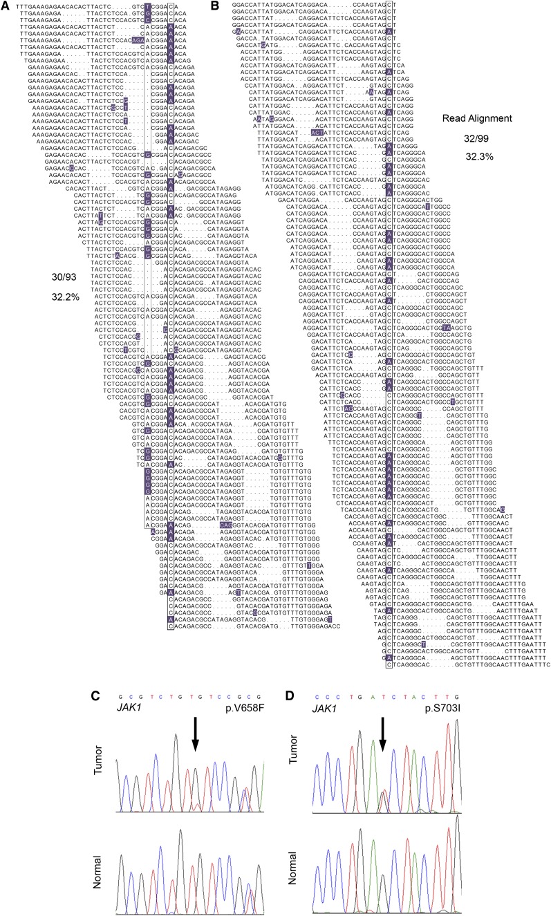
The comprehensive genetic alterations underlying the pathogenesis of T-cell prolymphocytic leukemia (T-PLL) are unknown. To address this, we performed whole-genome sequencing (WGS), whole-exome sequencing (WES), high-resolution copy-number analysis, and Sanger resequencing of a large cohort of T-PLL. WGS and WES identified novel mutations in recurrently altered genes not previously implicated in T-PLL including EZH2, FBXW10, and CHEK2. Strikingly, WGS and/or WES showed largely mutually exclusive mutations affecting IL2RG, JAK1, JAK3, or STAT5B in 38 of 50 T-PLL genomes (76.0%). Notably, gain-of-function IL2RG mutations are novel and have not been reported in any form of cancer. Further, high-frequency mutations in STAT5B have not been previously reported in T-PLL. Functionally, IL2RG-JAK1-JAK3-STAT5B mutations led to signal transducer and activator of transcription 5 (STAT5) hyperactivation, transformed Ba/F3 cells resulting in cytokine-independent growth, and/or enhanced colony formation in Jurkat T cells. Importantly, primary T-PLL cells exhibited constitutive activation of STAT5, and targeted pharmacologic inhibition of STAT5 with pimozide induced apoptosis in primary T-PLL cells. These results for the first time provide a portrait of the mutational landscape of T-PLL and implicate deregulation of DNA repair and epigenetic modulators as well as high-frequency mutational activation of the IL2RG-JAK1-JAK3-STAT5B axis in the pathogenesis of T-PLL. These findings offer opportunities for novel targeted therapies in this aggressive leukemia.


Figures

Figure 1
Figure 1
WGS identifies JAK1 mutations in 2 out of 4 index T-PLL cases. (A-B) The total individual reads supporting variant calling of the JAK1 p.V658F (A) and p.S703I (B) mutations in index T-PLL samples are shown. Nucleotides with a deviation from the reference sequence are highlighted. The mutations as well as a synonymous single-nucleotide polymorphism (A) are boxed. Dots in individual reads (A-B) represent unsequenced nucleotides (as opposed to sequence gaps) and are intrinsic to the self-assembling DNA nanoarray next-generation sequencing platform and unchained base-reads analysis approach used for WGS (see supplemental Methods for further details). (C-D) Sanger resequencing confirmation of the somatic acquisition of these mutations is shown.
Figure 2
Figure 2
High-frequency IL2RG-JAK1-JAK3-STAT5B mutations in T-PLL. (A-D) Representative IL2RG, JAK1, JAK3, and STAT5B mutations in primary T-PLL cells identified by WGS/WES and confirmed to be somatic by Sanger resequencing of tumor DNA (upper traces) and paired constitutional DNA (lower traces). (E) Schematic representations of mutations in IL2RG, JAK1, JAK3, and STAT5B identified through WGS, WES, or targeted Sanger resequencing of primary T-PLL (circles) and HUT78 cells (diamond). Confirmed somatic mutations are shown as filled symbols; variants where adequate matched constitutional DNA was not available are shown as open symbols. Mutations are clustered in the autoinhibitory pseudokinase domains of JAK1 and JAK3 (purple) or the SH2 domain of STAT5B (light blue) that mediates interactions between JAK and STAT proteins. One additional variant was detected in the kinase domain of JAK1 (red); a single case of T-PLL harbored a somatic 3-amino-acid deletion in the transmembrane domain of IL2RG (purple) as well as a somatic missense mutation in the cytoplasmic domain.
Figure 3
Figure 3
Three-dimensional localization of recurrent JAK3- and STAT5B-mutated amino acids. (A) The crystal structure of the SH2 domain of the STAT5A protein (1y1u) highlighting analogous residues of the STAT5B mutations p.N642H (blue), p.Y665H (purple), and p.T628S (red) demonstrating close 3-dimensional proximity of recurrently mutated STAT5B residues. Colored fill indicates an identical amino acid; white, minus indicates disparate residues; white with colored text, + indicates similar residues; and selected mutated residues are indicated in red. (B) The pseudokinase domain of JAK2 (4fvp) highlighting the V617 residue (light blue) recurrently mutated in myeloproliferative neoplasms and analogous residues for JAK3 mutations p.A573V (red), p.M511I (dark blue), and p.K563_C565del (purple). The extent of homology between the STAT5A and STAT5B or JAK2 and JAK3 in the regions of these recurrently mutated residues (red arrows) is highlighted below each respective 3-dimensional structure.
Figure 4
Figure 4
JAK-STAT mutations lead to increased pSTAT5 signaling, cytokine-independent growth, and enhanced colony formation. (A-B) Mutated IL2RG (p.G628_M630del), JAK1 (p.S703I), JAK3 (p.Q507P), and STAT5B (p.T628S and p.N642H) leads to increased activation of STAT5 transcriptional activity (A, bar graph; n = 3 for each mutant protein in separate experiments; asterisk indicates P < .05; dagger indicates P < .001) and increased phosphorylation of STAT5B (B, western blot; arrowhead indicates exogenous STAT5B, arrow indicates endogenous STAT5B; normalized densitometric pSTAT5/STAT5 ratios are indicated) in HeLa cells. STAT5B p.P267A represents a germline polymorphism. (C) Cytokine-independent cell proliferation in the presence of mutant p.T628S STAT5B protein in the cytokine-dependent Ba/F3 cell line cultured in the absence of growth factors (n = 6; asterisk indicates P < .01). (D) Enhanced colony-forming capacity of STAT5B p.N642H mutant in Jurkat T cells (n = 3; asterisk indicates P < .01).
Figure 5
Figure 5
Pimozide treatment of primary T-PLL cells to target increased pSTAT5B levels leads to reduced tumor cell growth and apoptosis. (A) Nuclear and cytoplasmic pY699 phosphorylated STAT5 in primary T-PLL samples by immunofluorescence microscopy (representative data are shown for 2 primary T-PLL cases [T-PLL25 and T-PLL38]). (B-C) Effects on HUT78 (pSTAT5-positive) viability following treatment with the JAK3 inhibitor tofacitinib (B; n = 3, P = .1144) and the STAT5 inhibitor pimozide (C; n = 3, asterisk indicates P < .0001). (D-F) Pharmacologic inhibition of pSTAT5 with pimozide leads to decreased viability (D) and diminished pSTAT5 levels (E) in primary T-PLL samples (n = 3 independent replicates for experiments in D; asterisks indicate P < .005; representative data are shown for T-PLL 25 in panel E). HH (pSTAT5 negative) is used as negative control, whereas SUDHL-1 (pSTAT5 positive) is used as a positive control. (F) A pathway diagram illustrates the interaction of IL2RG, JAK1, JAK3, and STAT5B during IL-2 cytokine activation. Cytokine binding to the extracellular portion of membrane-associated IL-2 receptors induces conformational change in the intracellular portion. Associated JAK nonreceptor tyrosine kinases autophosphorylate, leading to STAT recruitment and activation through tyrosine phosphorylation. Activated STAT proteins then dimerize and translocate to the nucleus to regulate transcription of numerous genes involved in differentiation, proliferation, and survival. Mutated components of the IL2R-JAK1-JAK3-STAT5B pathway are highlighted in green. Pimozide treatment inhibits STAT5B phosphorylation, limiting downstream transcriptional activation initiated by mutations in cytokine receptor/JAK-STAT proteins.

Comment in

References

    1. Matutes E, Brito-Babapulle V, Swansbury J, et al. Clinical and laboratory features of 78 cases of T-prolymphocytic leukemia. Blood. 1991;78(12):3269–3274. - PubMed
    1. Hopfinger G, Busch R, Pflug N, et al. Sequential chemoimmunotherapy of fludarabine, mitoxantrone, and cyclophosphamide induction followed by alemtuzumab consolidation is effective in T-cell prolymphocytic leukemia. Cancer. 2013;119(12):2258–2267. - PubMed
    1. Dearden CE. T-cell prolymphocytic leukemia. Clin Lymphoma Myeloma. 2009;(9)Suppl 3:S239-S243. - PubMed
    1. Virgilio L, Narducci MG, Isobe M, et al. Identification of the TCL1 gene involved in T-cell malignancies. Proc Natl Acad Sci USA. 1994;91(26):12530–12534. - PMC - PubMed
    1. Pekarsky Y, Hallas C, Isobe M, Russo G, Croce CM. Abnormalities at 14q32.1 in T cell malignancies involve two oncogenes. Proc Natl Acad Sci USA. 1999;96(6):2949–2951. - PMC - PubMed

Publication types

MeSH terms