

A neuroprotective brain-penetrating endopeptidase fusion protein ameliorates Alzheimer disease pathology and restores neurogenesis
- PMID: 24825898
- PMCID: PMC4067222
- DOI: 10.1074/jbc.M114.557439
A neuroprotective brain-penetrating endopeptidase fusion protein ameliorates Alzheimer disease pathology and restores neurogenesis
Abstract
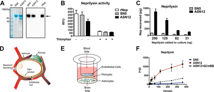
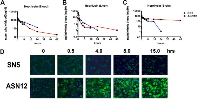
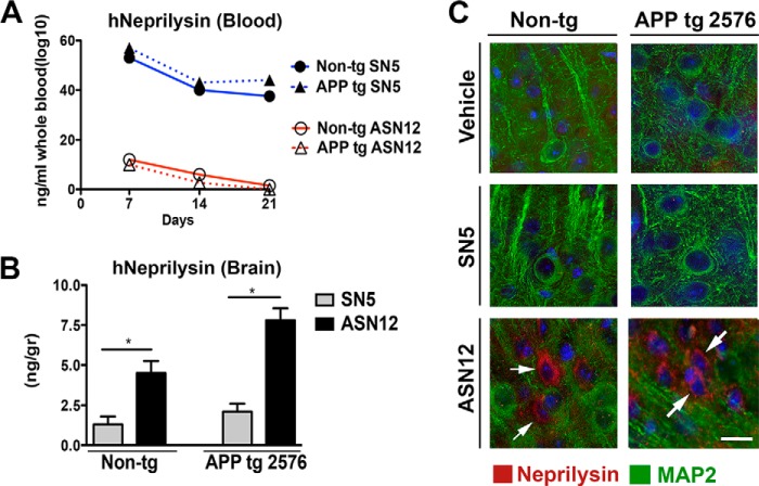
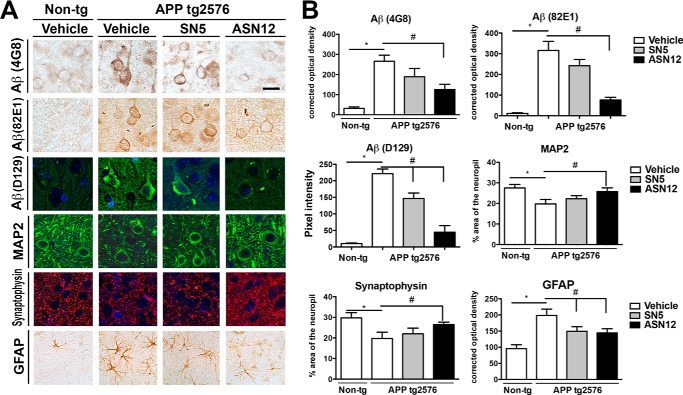
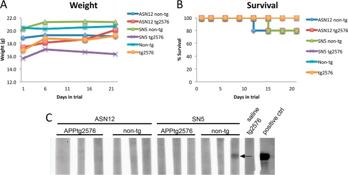
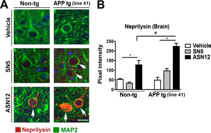
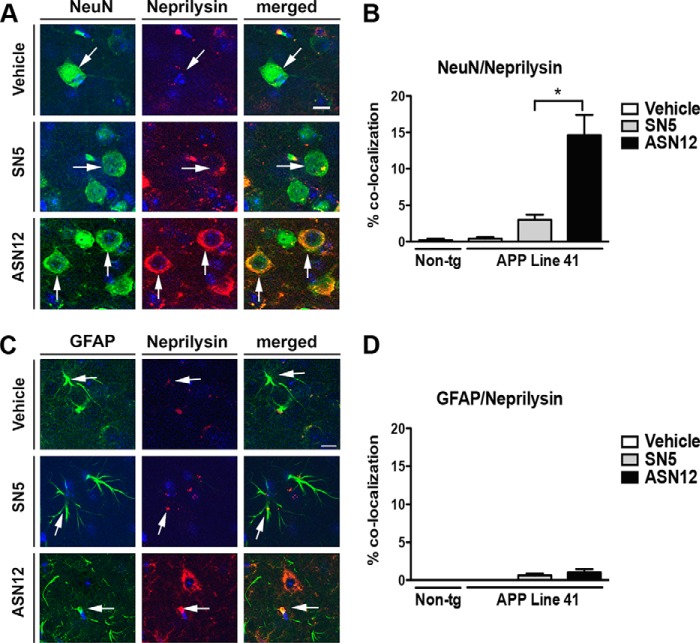
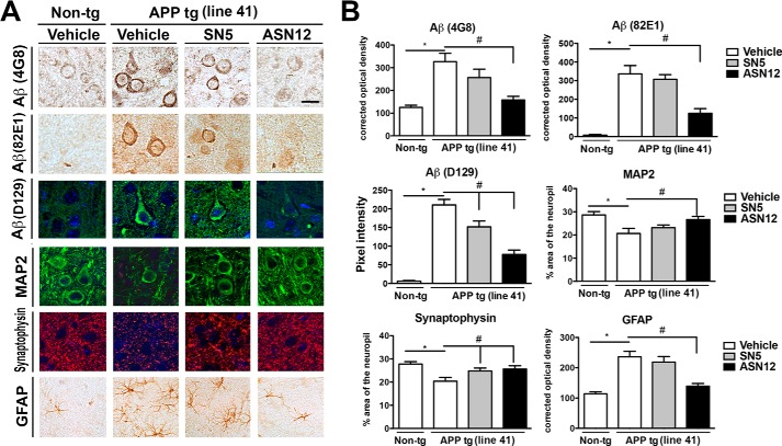
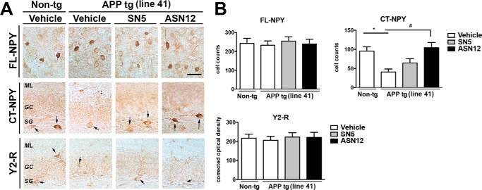
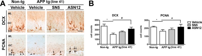
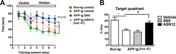
Alzheimer disease (AD) is characterized by widespread neurodegeneration throughout the association cortex and limbic system, deposition of amyloid-β peptide (Aβ) in the neuropil and around the blood vessels, and formation of neurofibrillary tangles. The endopeptidase neprilysin has been successfully used to reduce the accumulation of Aβ following intracranial viral vector delivery or ex vivo manipulated intracranial delivery. These therapies have relied on direct injections into the brain, whereas a clinically desirable therapy would involve i.v. infusion of a recombinant enzyme. We previously characterized a recombinant neprilysin that contained a 38-amino acid brain-targeting domain. Recombinant cell lines have been generated expressing this brain-targeted enzyme (ASN12). In this report, we characterize the ASN12 recombinant protein for pharmacology in a mouse as well as efficacy in two APPtg mouse models of AD. The recombinant ASN12 transited to the brain with a t½ of 24 h and accumulated to 1.7% of injected dose at 24 h following i.v. delivery. We examined pharmacodynamics in the tg2576 APPtg mouse with the prion promoter APP695 SWE mutation and in the Line41 mThy1 APP751 mutation mouse. Treatment of either APPtg mouse resulted in reduced Aβ, increased neuronal synapses, and improved learning and memory. In addition, the Line41 APPtg mice showed increased levels of C-terminal neuropeptide Y fragments and increased neurogenesis. These results suggest that the recombinant brain-targeted neprilysin, ASN12, may be an effective treatment for AD and warrant further investigation in clinical trials.
Keywords: Alzheimer Disease; Aβ; Blood Brain Barrier; Brain Penetrating Peptides; Lipoprotein Receptor; NPY; Neprilysin; Neurogenesis; Protein Targeting; Transcytosis.
© 2014 by The American Society for Biochemistry and Molecular Biology, Inc.
Figures
Similar articles
-
Systemic Central Nervous System (CNS)-targeted Delivery of Neuropeptide Y (NPY) Reduces Neurodegeneration and Increases Neural Precursor Cell Proliferation in a Mouse Model of Alzheimer Disease.J Biol Chem. 2016 Jan 22;291(4):1905-1920. doi: 10.1074/jbc.M115.678185. Epub 2015 Nov 30. J Biol Chem. 2016. PMID: 26620558 Free PMC article.
-
Enhanced neprilysin-mediated degradation of hippocampal Aβ42 with a somatostatin peptide that enters the brain.Theranostics. 2021 Jan 1;11(2):789-804. doi: 10.7150/thno.50263. eCollection 2021. Theranostics. 2021. PMID: 33391505 Free PMC article.
-
Neuropeptide Y fragments derived from neprilysin processing are neuroprotective in a transgenic model of Alzheimer's disease.J Neurosci. 2009 Jan 28;29(4):1115-25. doi: 10.1523/JNEUROSCI.4220-08.2009. J Neurosci. 2009. PMID: 19176820 Free PMC article.
-
APP transgenic modeling of Alzheimer's disease: mechanisms of neurodegeneration and aberrant neurogenesis.Brain Struct Funct. 2010 Mar;214(2-3):111-26. doi: 10.1007/s00429-009-0232-6. Epub 2009 Nov 29. Brain Struct Funct. 2010. PMID: 20091183 Free PMC article. Review.
-
Neprilysin gene transfer: A promising therapeutic approach for Alzheimer's disease.J Neurosci Res. 2015 Sep;93(9):1325-9. doi: 10.1002/jnr.23564. Epub 2015 Jun 11. J Neurosci Res. 2015. PMID: 26096375 Review.
Cited by
-
Aβ-Degrading Proteases: Therapeutic Potential in Alzheimer Disease.CNS Drugs. 2016 Aug;30(8):667-75. doi: 10.1007/s40263-016-0364-1. CNS Drugs. 2016. PMID: 27349988 Free PMC article. Review.
-
Applications of ApoB LDLR-Binding Domain Approach for the Development of CNS-Penetrating Peptides for Alzheimer's Disease.Methods Mol Biol. 2015;1324:331-7. doi: 10.1007/978-1-4939-2806-4_21. Methods Mol Biol. 2015. PMID: 26202279 Free PMC article.
-
A conformation-specific antibody against oligomeric β-amyloid restores neuronal integrity in a mouse model of Alzheimer's disease.J Biol Chem. 2021 Jan-Jun;296:100241. doi: 10.1074/jbc.RA120.015327. Epub 2021 Jan 9. J Biol Chem. 2021. PMID: 33376140 Free PMC article.
-
Brain-targeted stem cell gene therapy corrects mucopolysaccharidosis type II via multiple mechanisms.EMBO Mol Med. 2018 Jul;10(7):e8730. doi: 10.15252/emmm.201708730. EMBO Mol Med. 2018. PMID: 29884617 Free PMC article.
-
New Insights into Epigenetic and Pharmacological Regulation of Amyloid-Degrading Enzymes.Neurochem Res. 2016 Mar;41(3):620-30. doi: 10.1007/s11064-015-1703-1. Epub 2015 Sep 16. Neurochem Res. 2016. PMID: 26376806
References
-
- Ashford J. W. (2004) APOE genotype effects on Alzheimer's disease onset and epidemiology. J. Mol. Neurosci. 23, 157–165 - PubMed
-
- Terry R., Hansen L., Masliah E. (1994) in Alzheimer disease (Terry R., Katzman R., eds) pp. 179–196, Raven Press, New York
-
- Marr R. A., Spencer B. J. (2010) Diabetes NEP-like endopeptidases and Alzheimer's disease. Curr. Alzheimer. Res. 7, 223–229 - PubMed
-
- Howell S., Nalbantoglu J., Crine P. (1995) Neutral endopeptidase can hydrolyze β-amyloid(1–40) but shows no effect on β-amyloid precursor protein metabolism. Peptides 16, 647–652 - PubMed
-
- Iwata N., Tsubuki S., Takaki Y., Shirotani K., Lu B., Gerard N. P., Gerard C., Hama E., Lee H. J., Saido T. C. (2001) Metabolic regulation of brain Aβ by neprilysin. Science 292, 1550–1552 - PubMed
Publication types
MeSH terms
Substances
Grants and funding
LinkOut - more resources
Full Text Sources
Other Literature Sources
Medical
Molecular Biology Databases
Miscellaneous
