. 2014 Jun 27;289(26):18535-48.
doi: 10.1074/jbc.M114.569087. Epub 2014 May 13.

Mouse myosin-19 is a plus-end-directed, high-duty ratio molecular motor

Affiliations

Mouse myosin-19 is a plus-end-directed, high-duty ratio molecular motor

Zekuan Lu et al. J Biol Chem. .

Abstract

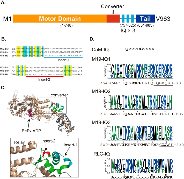
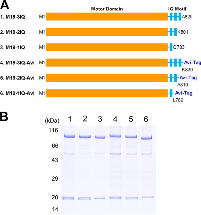
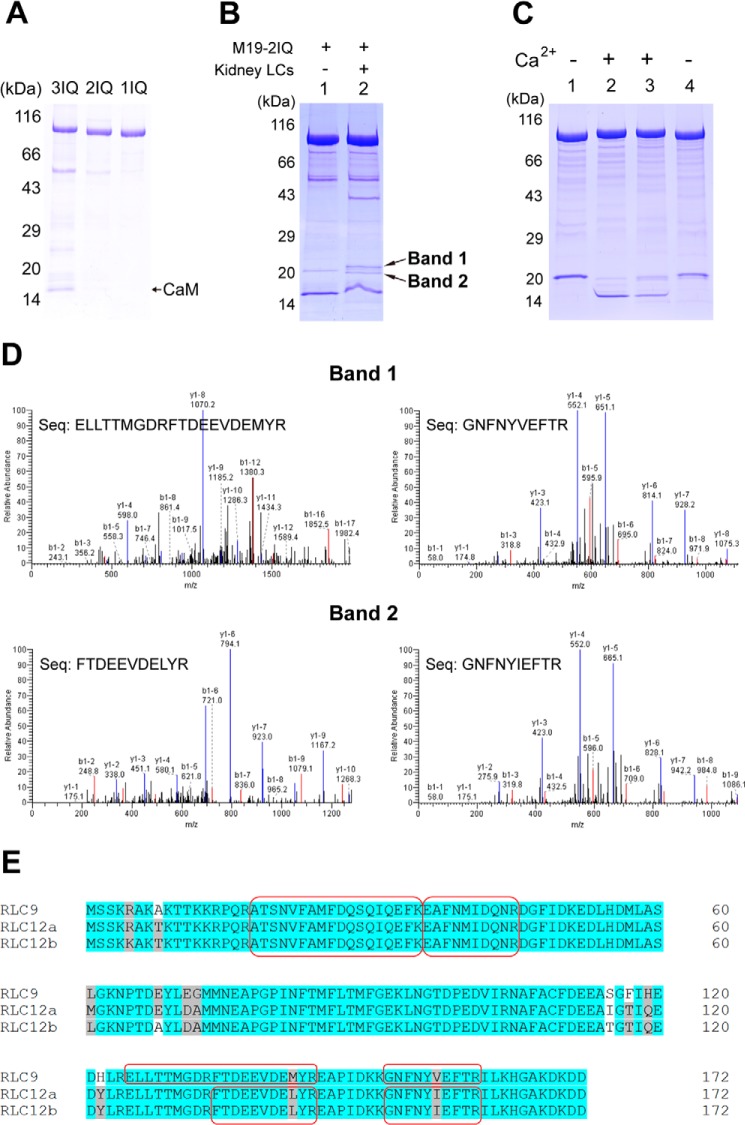
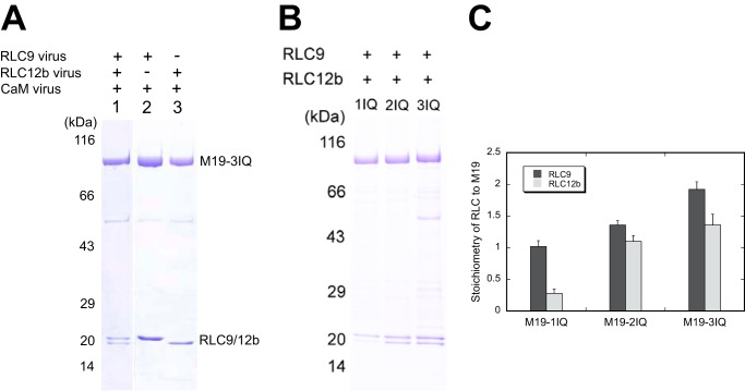
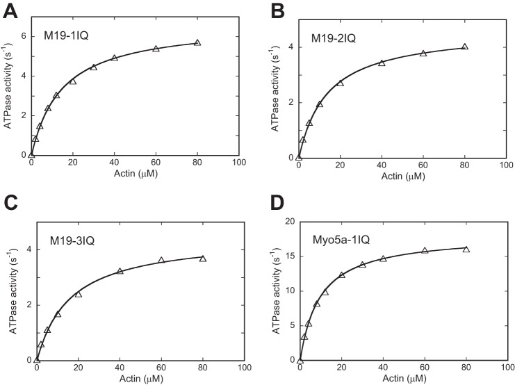
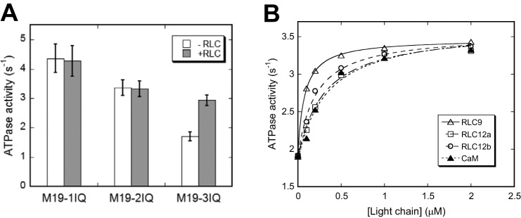
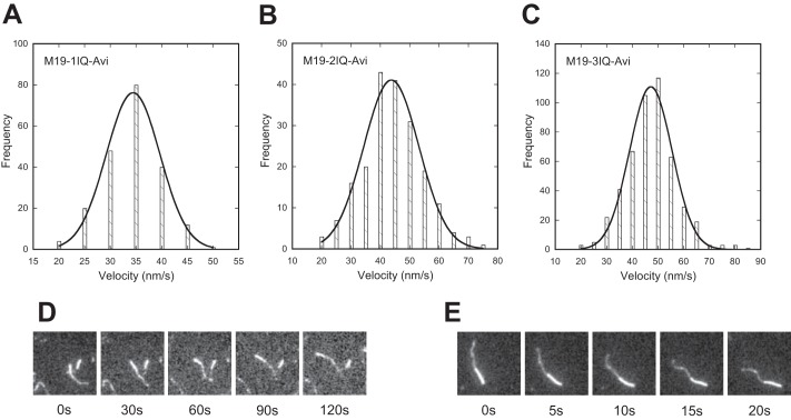
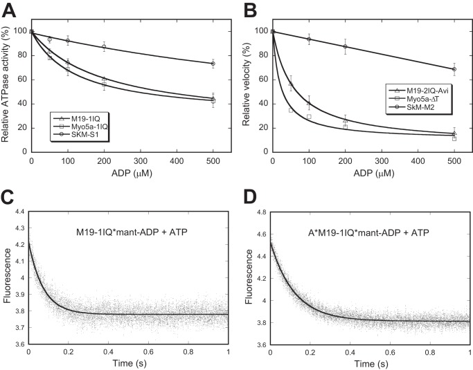
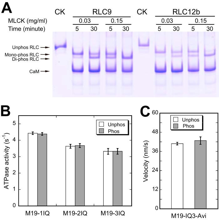
Class XIX myosin (Myo19) is a vertebrate-specific unconventional myosin, responsible for the transport of mitochondria. To characterize biochemical properties of Myo19, we prepared recombinant mouse Myo19-truncated constructs containing the motor domain and the IQ motifs using the baculovirus/Sf9 expression system. We identified regulatory light chain (RLC) of smooth muscle/non-muscle myosin-2 as the light chain of Myo19. The actin-activated ATPase activity and the actin-gliding velocity of Myo19-truncated constructs were about one-third and one-sixth as those of myosin-5a, respectively. The apparent affinity of Myo19 to actin was about the same as that of myosin-5a. The RLCs bound to Myo19 could be phosphorylated by myosin light chain kinase, but this phosphorylation had little effect on the actin-activated ATPase activity and the actin-gliding activity of Myo19-truncated constructs. Using dual fluorescence-labeled actin filaments, we determined that Myo19 is a plus-end-directed molecular motor. We found that, similar to that of the high-duty ratio myosin, such as myosin-5a, ADP release rate was comparable with the maximal actin-activated ATPase activity of Myo19, indicating that ADP release is a rate-limiting step for the ATPase cycle of acto-Myo19. ADP strongly inhibited the actin-activated ATPase activity and actin-gliding activity of Myo19-truncated constructs. Based on the above results, we concluded that Myo19 is a high-duty ratio molecular motor moving to the plus-end of the actin filament.

Keywords: ATPase; Actin; Intracellular Trafficking; Mitochondria; Myosin.


Figures

FIGURE 1.
FIGURE 1.
Diagram of Myo19 structure and sequence alignment of Myo19. A, predicted structure of Myo19. The diagram was not drawn to scale. B, sequence alignment of the converter of myosin-5a and Myo19 reveals two inserts in the converter of Myo19. Yellow and blue shades indicate the identical and conserved residues, respectively. Myo5a-Mm, mouse myosin-5a (GI: 115511052); M19-Mm, mouse Myo19 (GI: 254939539); M19-Hs, human Myo19 (GI: 254939537). C, location of the two Myo19-specific inserts in the crystal structure of myosin-5a (PDB code 1W7J). Upper, the head domain of myosin-5a; lower, the expanded view of the converter. The converter is colored green with insert 1 in cyan and insert 2 in red. Insert 1 and 2 are adjacent to each other and insert 2 is adjacent to the tip of the relay helix. D, consensus sequences of CaM-binding IQ motif, IQ motifs of Myo19, and RLC-binding IQ motif of myosin-2 and -18. CaM-IQ, consensus sequence of the six IQ motifs of mouse myosin-5a (GI: 115511052). M19-IQ1, -IQ2, and -IQ3, sequence logo of the consensus sequence of the three IQ motifs of 11 class XIX myosins, including mouse (GI: 254939539), rat (GI: 254939541), human (GI: 254939537), cattle (GI: 254939551), elephant (GI: 344285323), rabbit (GI: 291405654), cat (GI: 410980571), whale (GI: 466018728), chicken (GI: 513216545), Xenopus laevis (GI: 82178330), and zebrafish (GI: 189519181). The sequence shown at the bottom is mouse Myo19. Conserved residues are shown in bold. Underlined residues are not essential for RLC binding (for details see ”Discussion“). RLC-IQ, sequence logo of the consensus sequence of IQ2 motifs of five muscle myosin-2 and two myosin-18, including scallop muscle myosin (GI: 5611), human non-muscle myosin-2a (GI: 47678583), -2b (GI: 219841954), -2c (GI: 116284394), smooth muscle myosin (GI: 46486992), myosin-18a (GI: 24660442), and myosin-18b (GI: 219841774). Sequence logos were generated by an online program, WebLOGO.
FIGURE 2.
FIGURE 2.
Myo19 truncated constructs. A, schematic diagrams of Myo19 constructs used in this study. To facilitate purification, a FLAG tag was attached to the N terminus of each construct. To facilitate actin-gliding assay, an Avi-tag was attached to the C terminus of the last three constructs. Amino acid numbers of the constructs are indicated. B, SDS-PAGE (4–20%) of purified Myo19-truncated constructs. Myo19-truncated construct was coexpressed with RLC9 and RLC12b in Sf9 cells and purified by anti-FLAG affinity agarose. Lane 1, M19-3IQ; 2, M19-2IQ; 3, M19-1IQ; 4, M19-3IQ-Avi; 5, M19-2IQ-Avi; 6, M19-1IQ-Avi.
FIGURE 3.
FIGURE 3.
Identification of the light chain of Myo19. A, SDS-PAGE (4–20%) of the purified Myo19-truncated constructs coexpressed with CaM in Sf9 cells. B, SDS-PAGE (4–20%) of M19-2IQ co-purified with crude extract of myosin light chain from mouse kidney. Lane 1, purified M19-2IQ expressed in Sf9 cells; 2, re-purified M19-2IQ after incubated with crude extract of myosin light chain from mouse kidney. Band-1 and band-2 indicate the two specific ∼20-kDa bands co-purified with M19-2IQ. C, CaM gel shift assay of the light chain co-purified with M19-2IQ. Lanes 1 and 2, purified M19-2IQ expressed in Sf9 cells; lanes 3 and 4, re-purified M19-2IQ after incubation with crude extract of myosin light chain from mouse kidney. Lanes 1 and 4 were run under EGTA conditions, lanes 2 and 3 were run under Ca2+ conditions. For details, see ”Experimental Procedures.“ D, MS/MS analysis of band-1 and band-2 in lane 2, the two specific ∼20-kDa bands co-purified with M19-2IQ. Two peptides corresponding to RLC9 and RLC12b were detected in band-1 and band-2, respectively. The sequences of the peptides were shown. E, amino acid sequence alignment of three isoforms of mouse RLC. The sequences shaded in gray are conserved but not identical residues, and the ones in white are non-conserved residues. The peptide sequences identified in MS/MS are boxed.
FIGURE 4.
FIGURE 4.
Identification of the light chain binding sites in Myo19. Myo19-truncated constructs were co-expressed with different groups of light chains. The purified Myo19 samples were subjected to SDS-PAGE (4–20%) and Coomassie Blue staining. A, SDS-PAGE of purified M19-3IQ coexpressed with the different light chains (indicated at the top of the gel) in Sf9 cells. B, SDS-PAGE of purified M19-1IQ, -2IQ, and -3IQ coexpressed with RLC9 and RLC12b. C, stoichiometry of RLC9 and RLC12b to Myo19 heavy chain. Quantifications of the light chain and heavy chain were done with ImageJ software. The molar ratio of RLC versus the heavy chain was calculated based on the corresponding molecular weight masses. Values are mean ± S.D. from three independent assays.
FIGURE 5.
FIGURE 5.
Actin-activated ATPase activities of Myo19-truncated constructs. ATPase activities of M19-1IQ (A), M19-2IQ (B), and M19-3IQ (C) were measured in a solution containing 50 mm NaCl, 20 mm MOPS (pH 7.0), 1 mm MgCl2, 1 mm EGTA, 0.25 mg/ml of BSA, 1 mm DTT, 0.5 mm ATP, 2.5 mm phosphoenol pyruvate, 20 units/ml of pyruvate kinase, 4 μm RLC9, 4 μm RLC12b, and 0–80 μm actin. ATPase activity of Myo5a-1IQ (D) was measured similarly except that RLCs was substituted with 12 μm CaM. Curves are the least squares fits of the data points based upon the equation, V = (Vmax × [actin]/Kactin + [actin]). Although multiple independent assays of at least 2 preparations have been performed, data from a single assay are presented. Vmax and Kactin from multiple assays are summarized in Table 1.
FIGURE 6.
FIGURE 6.
Exogenous light chain enhances actin-activated ATPase activity of M19–3IQ. A, actin-activated ATPase activity of Myo19-truncated constructs in the presence or absence of 4 μm RLC9 and RLC12b. Values are mean ± S.D. from two to four independent assays. B, effects of exogenous light chain on actin-activated ATPase activity of M19-3IQ. ATPase assay was performed in 50 mm NaCl, 20 mm MOPS (pH 7.0), 1 mm MgCl2, 1 mm EGTA, 0.25 mg/ml of BSA, 1 mm DTT, 0.5 mm ATP, 2.5 mm phosphoenol pyruvate, 20 units/ml of pyruvate kinase, 40 μm actin, and 0–2 μm light chain. The stimulation of ATPase activities by exogenous light chain was fitted with a hyperbolic equation: V = V0 + Vmax × [LC]/(Kd + [LC]), where V0, the activity in the absence of exogenous light chain; Vmax, the activity stimulated by exogenous light chain; [LC], the concentration of exogenous light chain; Kd, the apparent affinity between the light chain and M19-IQ3. The apparent Kd values are 0.076 ± 0.027, 0.321 ± 0.076, 0.223 ± 0.068, and 0.335 ± 0.049 μm for RLC9, RLC12a, RLC12b, and CaM, respectively. Data were average of two independent assays.
FIGURE 7.
FIGURE 7.
Actin-gliding activity of Myo19. Biotinylated Myo19-truncated constructs were attached to a coverslip via biotinylated BSA and streptavidin, and the movements of rhodamine-labeled actin filaments were recorded with an Olympus IX71 inverted microscope at room temperature (about 25 °C). The velocity was analyzed by MATLAB. The solid line shows a fit to a single Gaussian curve. For details, see ”Experimental Procedures.“ A–C, histogram of multimolecular actin-gliding velocity of Myo19-truncated constructs, i.e. M19-1IQ-Avi (A), M19-2IQ-Avi (B), and M19-3IQ-Avi (C). D and E, direction of the actin filament translocated by M19-1IQ-Avi (D) and Myo5a-ΔT. E, actin filaments were dual fluorescence-labeled. Plus-end was labeled with Alexa 488 (dim) and minus end with rhodamine (bright). Actin filaments were moved toward the minus-end by M19-1IQ-Avi and Myo5a-ΔT.
FIGURE 8.
FIGURE 8.
ADP-off is the rate-limiting step for the motor activity of Myo19. A, effects of ADP on actin-activated ATPase activity of M19-1IQ, Myo5a-1IQ, and SkM-S1. Actin-activated ATPase activity was measured in the presence of 40 μm actin, 500 μm ATP, and 0–500 μm ADP and the released phosphate was determined by the malachite green method. The inhibition of ATPase activities by ADP was fitted with a hypophobic equation. B, effects of ADP on actin-gliding activity of M19–1IQ, Myo5a-ΔT, and skeletal muscle myosin (SkM-M2). Actin-gliding activity was measured in the presence of 500 μm ATP and 0–500 μm ADP. In the absence of ADP, the actin-gliding activities of M19-2IQ-Avi, Myo5a-ΔT, and skeletal muscle myosin were 40, 280, and 3.64 μm/s, respectively. C and D, dissociation of mant-ADP from M19-1IQ (C) and acto-M19-1IQ (D). The fluorescence intensity of mant-ADP was recorded in a stopped flow after mixing 1 mm ATP with and 1 μm mant-ADP and 1 μm M19–1IQ in the absence (C) or presence (D) of 1.5 μm phalloidin-stabilized actin filaments. The smooth line is the fit to single exponential kinetics, with Kobs of 13.65 s−1 (C) and 8.37 s−1 (D).
FIGURE 9.
FIGURE 9.
Effects of RLC phosphorylation on the motor function of Myo19. A, phosphorylation of the RLC of M19-3IQ by MLCK. M19-3IQ was co-expressed with RLC9 or RLC12b in Sf9 cells and purified by anti-FLAG affinity chromatography. The purified M19-3IQ/RLC9 or M19-3IQ/RLC12b was incubated with MLCK (0.03 mg/ml) in phosphorylation solution (30 mm Tris-HCl, pH 7.5, 50 mm KCl, 1 mm DTT, 1 mm MgCl2, 10 μm CaM, 0.1 mm CaCl2, and 1 mm ATP) at 25 °C for the indicated times. Phosphorylation of RLC was determined by urea/glycerol PAGE and visualized by Coomassie Blue staining. CK represents the unphosphorylated control, which was not treated with MLCK. B, effects of RLC phosphorylation on the actin-activated ATPase activity of Myo19. ATPase assay was performed in 50 mm KCl, 25 mm Tris-HCl (pH 7.5), 1 mm MgCl2, 0.25 mg/ml of BSA, 1 mm DTT, 0.5 mm ATP, 2.5 mm phosphoenol pyruvate, 20 units/ml of pyruvate kinase, 40 μm actin, 0.1 mm CaCl2, 2 μm CaM with 0.03 mg/mg of MLCK (phosphorylated conditions) or without MLCK (unphosphorylated conditions) at 25 °C. The reaction was stopped at various times between 4 and 60 min as described under “Experimental Procedures.” C, effects of RLC phosphorylation on in vitro actin-gliding activity of Myo19-3IQ-Avi. Prior to actin-gliding assay, biotinylated M19-3IQ-Avi was phosphorylated by 0.15 mg/ml of MLCK in phosphorylation solution at 25 °C for 5 min. Actin-gliding assay was performed as described in the legend of Fig. 7.

Similar articles

Cited by

References

    1. Hartman M. A., Finan D., Sivaramakrishnan S., Spudich J. A. (2011) Principles of unconventional myosin function and targeting. Annu. Rev. Cell Dev. Biol. 27, 133–155 - PMC - PubMed
    1. Hammer J. A., 3rd, Sellers J. R. (2012) Walking to work: roles for class V myosins as cargo transporters. Nat. Rev. Mol. Cell Biol. 13, 13–26 - PubMed
    1. Akhmanova A., Hammer J. A., 3rd (2010) Linking molecular motors to membrane cargo. Curr. Opin. Cell Biol. 22, 479–487 - PMC - PubMed
    1. Foth B. J., Goedecke M. C., Soldati D. (2006) From the cover: new insights into myosin evolution and classification. Proc. Natl. Acad. Sci. U.S.A. 103, 3681–3686 - PMC - PubMed
    1. Odronitz F., Kollmar M. (2007) Drawing the tree of eukaryotic life based on the analysis of 2,269 manually annotated myosins from 328 species. Genome Biol. 8, R196. - PMC - PubMed

Publication types

Associated data