

Incidental discovery of a membranous ventricular septal aneurysm in two dissimilar patients
- PMID: 24826245
- PMCID: PMC4008397
- DOI: 10.1155/2012/324326
Incidental discovery of a membranous ventricular septal aneurysm in two dissimilar patients
Abstract
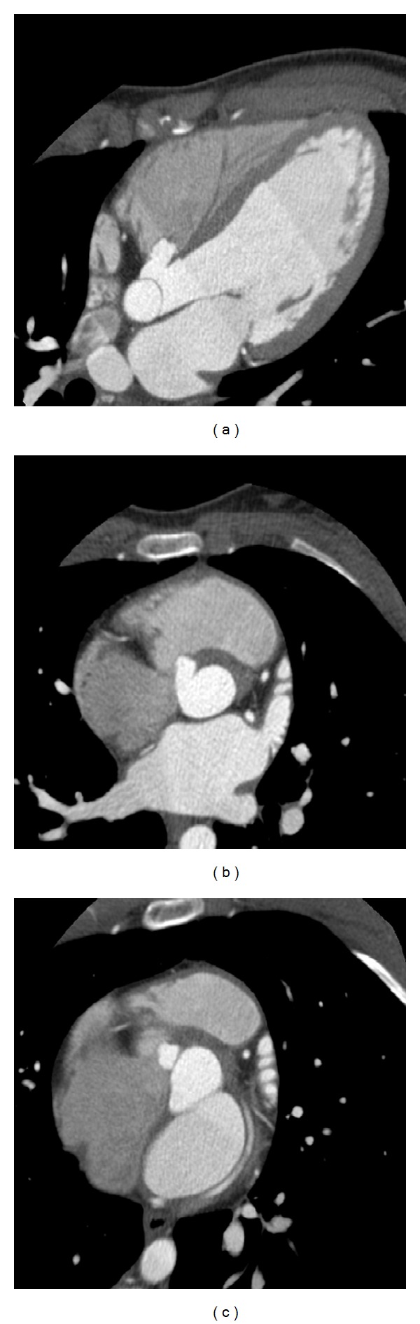
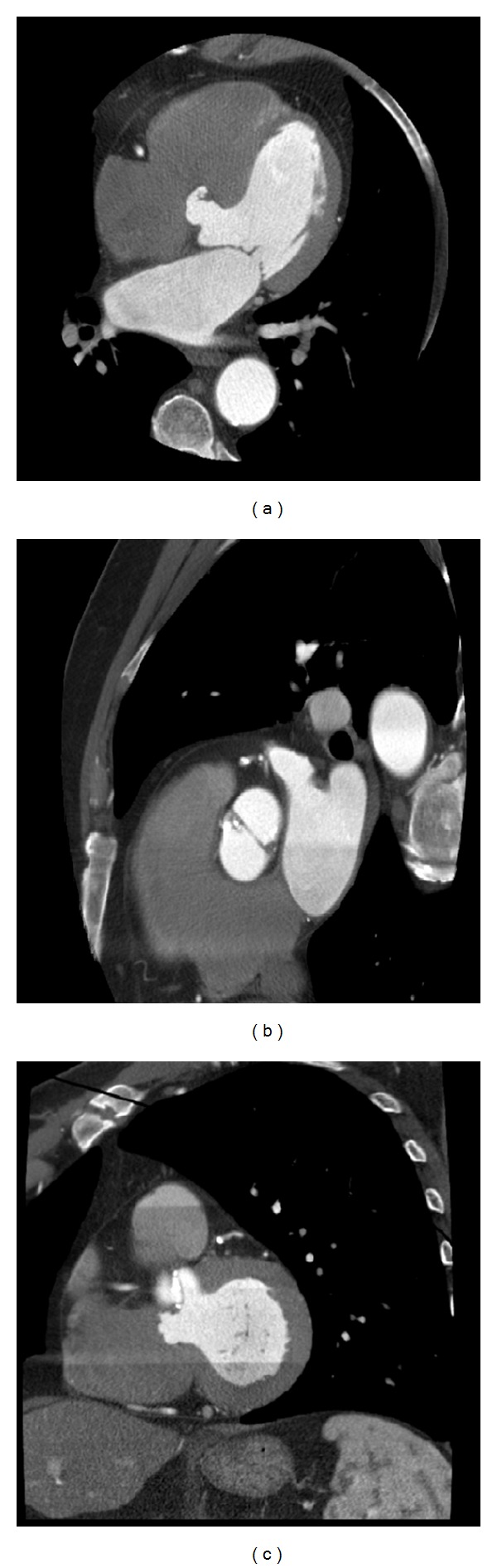
A ventricular septal aneurysm (VSA) is a rare cardiac anomaly, and an accurate statistic of its prevalence has not been reported in the literature. True incidence is likely underestimated as most patients are thought to be asymptomatic. As a result, most VSAs are discovered incidentally on echocardiography, during angiography, or at autopsy. Potential complications include rupture, bacterial endocarditis, right ventricular outflow tract obstruction, and thromboembolic disease. It has been proposed that VSAs occur in association with ventricular septal defects (VSDs) and other congenital cardiac abnormalities. It is uncommon for a VSA to exist in the absence of a known prior ventricular septal defect. We present two cases, each highlighting an incidental intact aneurysm involving the membranous interventricular septum. We discuss the contrast in the two patients with regard to their age, accompanying cardiac anomalies and cardiovascular fitness. Clinical implications of the condition are reviewed.
Figures
References
-
- Choi M, Jung JI, et al. Ventricular septal aneurysms in adults: findings of Cardiac CT images and correlation with clinical features. Acta Radiologica. 2011;52:619–623. - PubMed
-
- Yavuz S, Eris C, Goncu T, Sezen M, Ata Y, Turk T. An incidental aneurysm of the interventricular membranous septum. Archives of Iranian Medicine. 2010;13(4):363–364. - PubMed
-
- Yilmaz AT, Özal E, Arslan M, Tatar H, Öztürk ÖY. Aneurysm of the membranous septum in adult patients with perimembranous ventricular septal defect. European Journal of Cardio-thoracic Surgery. 1997;11(2):307–311. - PubMed
-
- Espinoza J, Kalache K, Gonçalves LF, et al. Prenatal diagnosis of membranous ventricular septal aneurysms and their association with absence of atrioventricular valve ‘offsetting’. Ultrasound in Obstetrics and Gynecology. 2004;24(7):787–792. - PubMed
LinkOut - more resources
Full Text Sources
Research Materials
