Bilateral absence of the superior vena cava
- PMID: 24826253
- PMCID: PMC4008471
- DOI: 10.1155/2012/461040
Bilateral absence of the superior vena cava
Abstract
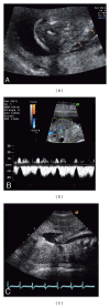
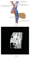
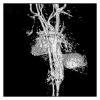
Bilateral absence of the superior vena cava (SVC) is a very rarely detected, mainly asymptomatic congenital vascular anomaly. Though usually innocent, this anomaly may complicate cardiothoracic surgery and certain procedures like central venous catheter insertion. This SVC anomaly is poorly known, and we assume that its incidence in the general population may be higher than detected. In this paper, we summarize current knowledge on this anomaly and its clinical implications. In addition, we present a neonatal case with bilateral absence of the SVC associated with a fetal cystic hygroma. Conclusion. Totally absent SVC can cause unexpected problems during cardiothoracic surgery. Suspicion of SVC absence should arise in basic echocardiography. Our paper suggests that, like other congenital anomalies, bilateral absent SVC may be associated with a fetal cyctic hygroma.
Figures


References
-
- Bartram U, Van Praagh S, Levine JC, Hines M, Bensky AS, Van Praagh R. Absent right superior vena cava in visceroatrial situs solitus. American Journal of Cardiology. 1997;80(2):175–183. - PubMed
-
- Hussain SA, Chakravarty S, Chaikhouni A, Smith JR. Congenital absence of superior vena cava: unusual anomaly of superior systemic veins complicating pacemaker placement. PACE. 1981;4(3):328–334. - PubMed
-
- Del Ojo JL, Jimenez CD, Vilches PJ, Serrano AL, Gutierrez FL, Rodriguez JB. Absence of the superior vena cava: difficulties for pacemaker implantation. PACE. 1999;22(7):1103–1105. - PubMed
-
- Saunders RN, Richens DR, Morris GK. Bilateral absence of the superior vena cava. Annals of Thoracic Surgery. 2001;71(6):2041–2043. - PubMed
-
- Minniti S, Visentini S, Procacci C. Congenital anomalies of the venac cavae: embryological origin, imaging features and report of three new variants. European Radiology. 2002;12(8):2040–2055. - PubMed
LinkOut - more resources
Full Text Sources

