Multiple multilateral coronary-cameral fistulae in a patient with minor cardiac venous system
- PMID: 24826312
- PMCID: PMC4008345
- DOI: 10.1155/2014/754703
Multiple multilateral coronary-cameral fistulae in a patient with minor cardiac venous system
Abstract
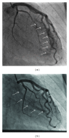
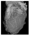
A 40-year-old man was hospitalized in the coronary care unit with chest pain and abnormal electrocardiogram. Twenty days earlier, the patient underwent laparoscopic gallbladder surgery. Due to chest pain and ischemic ECG changes, patient was subjected to coronary angiography. The selective coronary angiography revealed multiple multilateral fistulae arising from the left anterior descending artery, circumflex artery, and the right coronary artery draining to the left ventricle. Multislice computed tomography showed hypoplastic coronary sinus and minor cardiac venous system.
Figures


Similar articles
-
Bilateral Coronary Pulmonary Artery Fistulae.J Assoc Physicians India. 2016 May;64(5):77. J Assoc Physicians India. 2016. PMID: 27735159
-
A Coronary Cameral Fistula Draining Into Left Ventricle: A Rare Finding.Cureus. 2022 Jun 8;14(6):e25755. doi: 10.7759/cureus.25755. eCollection 2022 Jun. Cureus. 2022. PMID: 35812593 Free PMC article.
-
A rare case of dual coronary cameral fistulae.Clin Case Rep. 2023 Dec 9;11(12):e8300. doi: 10.1002/ccr3.8300. eCollection 2023 Dec. Clin Case Rep. 2023. PMID: 38084354 Free PMC article.
-
Unilateral and multilateral congenital coronary-pulmonary fistulas in adults: clinical presentation, diagnostic modalities, and management with a brief review of the literature.Clin Cardiol. 2014 Sep;37(9):536-45. doi: 10.1002/clc.22297. Epub 2014 Sep 5. Clin Cardiol. 2014. PMID: 25196980 Free PMC article. Review.
-
A rare coronary anomaly consisting of a single right coronary ostium in an adult undergoing surgical coronary revascularization: a case report and review of the literature.J Med Case Rep. 2016 Jul 1;10(1):190. doi: 10.1186/s13256-016-0977-5. J Med Case Rep. 2016. PMID: 27370010 Free PMC article. Review.
References
-
- Yamanaka O, Hobbs RE. Coronary artery anomalies in 126,595 patients undergoing coronary arteriography. Catheterization and Cardiovascular Diagnosis. 1990;21(1):28–40. - PubMed
-
- Gillebert C, van Hoof R, van de Werf F, Piessens J, de Geest H. Coronary artery fistulas in an adult population. European Heart Journal. 1986;7(5):437–443. - PubMed
-
- Luo L, Kebede S, Wu S, Stouffer GA. Coronary artery fistulae. The American Journal of the Medical Sciences. 2006;332(2):79–84. - PubMed
-
- Black IW, Loo CK, Allan RM. Multiple coronary artery-left ventricular fistulae: clinical, angiographic, and pathologic findings. Catheterization and Cardiovascular Diagnosis. 1991;23(2):133–135. - PubMed
LinkOut - more resources
Full Text Sources
Other Literature Sources

