. 2014 May 14;9(5):e97257.
doi: 10.1371/journal.pone.0097257. eCollection 2014.

Dual role of novel ingenol derivatives from Euphorbia tirucalli in HIV replication: inhibition of de novo infection and activation of viral LTR

Affiliations

Dual role of novel ingenol derivatives from Euphorbia tirucalli in HIV replication: inhibition of de novo infection and activation of viral LTR

Celina M Abreu et al. PLoS One. .

Abstract

HIV infection is not cleared by antiretroviral drugs due to the presence of latently infected cells that are not eliminated with current therapies and persist in the blood and organs of infected patients. New compounds to activate these latent reservoirs have been evaluated so that, along with HAART, they can be used to activate latent virus and eliminate the latently infected cells resulting in eradication of viral infection. Here we describe three novel diterpenes isolated from the sap of Euphorbia tirucalli, a tropical shrub. These molecules, identified as ingenols, were modified at carbon 3 and termed ingenol synthetic derivatives (ISD). They activated the HIV-LTR in reporter cell lines and human PBMCs with latent virus in concentrations as low as 10 nM. ISDs were also able to inhibit the replication of HIV-1 subtype B and C in MT-4 cells and human PBMCs at concentrations of EC50 0.02 and 0.09 µM respectively, which are comparable to the EC50 of some antiretroviral currently used in AIDS treatment. Control of viral replication may be caused by downregulation of surface CD4, CCR5 and CXCR4 observed after ISD treatment in vitro. These compounds appear to be less cytotoxic than other diterpenes such as PMA and prostratin, with effective dose versus toxic dose TI>400. Although the mechanisms of action of the three ISDs are primarily attributed to the PKC pathway, downregulation of surface receptors and stimulation of the viral LTR might be differentially modulated by different PKC isoforms.


Conflict of interest statement

Competing Interests: LFP is a share holder of KyoLab Laboratories. He also has a contract with Amazonia Fitomedicamentos to develop the ingenol derivatives. This does not alter the authors' adherence to PLOS ONE policies on sharing data and materials.

Figures

Figure 1
Figure 1. Chemical structures of ingenol synthetic derivatives indicating the modifications at carbon 3.
A) Ingenol core; B) Ingenol-3-trans-cinnamate (ING-A); C) Ingenol-3-hexanoate (ING-B); D) Ingenol-3-dodecanoate (ING-C).
Figure 2
Figure 2. ISDs upregulate HIV-LTR-driven GFP expression in J-Lat clones.
GFP expression (A) and viability (B) in J-Lat clone 9.2 were measured after cells were treated with ISDs, prostratin and PMA in different concentrations for 48 h. Similar experiment was done in J-Lat clone 10.6 (C and D). E) Comparison of GFP expression in four different J-Lat clones, all treated with 1 µM of each compound for 48 h. J-Lat clone 15.4 is depicted in a different graph (F). Graphs show mean and standard deviation of 3 separate tests. In all experiments, TNF-α 10 ng/mL was used as positive control.
Figure 3
Figure 3. ISDs upregulate the production of HIV particles in diverse cell models.
A) Four J-Lat clones were treated with 1 µM of different diterpenes and p24 levels in the supernatant were quantitated by ELISA. Similar experiment was done in J-Lat clone 15.4 (B) and other latent cell models (C). Graphs show mean and standard deviation of 3 separate tests. In all experiments, TNF-α 10 ng/mL was used as positive control.
Figure 4
Figure 4. ISDs upregulate HIV-LTR-driven luciferase expression in human PBMCs.
Blood-derived PBMCs from three healthy donors were transfected with pLTR-luc and then treated with different concentrations of ISDs, prostratin and PMA for 24 and 72 h. Graphs show levels of luciferase activity after 24 h (A) and cell viability after 24 (B) and 72 h (C). Results are represented as mean plus standard deviation of three independent experiments. DMSO 1% was used as vehicle control (untreated – yellow bar).
Figure 5
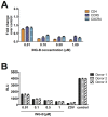
Figure 5. ISDs reduce HIV replication in a dose-dependent manner.
Lymphocytic cell line MT-4 (A and B) and human PBMCs (C and D) were infected with HIV subtype B NL-4.3 (A and C) or HIV subtype C ZM247Fv-1 (B and D) and treated with different concentrations of ISDs. After six days, cell viability was evaluated using the Cell Titer blue kit. Curves were derived by non-linear regression (dose-response curve by Hill for 3 parameters), and dotted lines represent EC50. Experiments were done in triplicate for PBMCs and sextuplicate for MT-4. ZVD was used as positive control.
Figure 6
Figure 6. ISDs modulate surface expression of CD4, CXCR4, CCR5 and activation markers.
MT-4 cells (A) and CD4+ T lymphocytes (B) were stimulated with ISDs (ING-A, ING-B, ING-C), bryostatin (bryo), prostratin (prost) and PMA for 48 h. Surface marker expression was evaluated by cytometry. CD4+ T cells were also evaluated for the activation markers CD25, CD38, HLA-DR (C) and CD69 (D). Results are representative of the mean and standard deviation of three independent experiments. DMSO 1% was used as vehicle control.
Figure 7
Figure 7. ING-B blocks HIV de novo replication through downregulation of surface receptors.
CD4+ T cells from three healthy donors were stimulated with PHA/IL-2 for 5 days and then treated with different concentrations of ING-B for 24 h. A portion of the cells was used for cytometry evaluation (A), and the graph depicts the mean and standard deviation of three independent experiments. The remaining cells were infected with HIV NL4-3-Luc for 24 h and cell lysates were analyzed by luciferase activity (B). Results are shown as relative light units (RLU). ZVD 1 µM was used as positive control and DMSO 1% as vehicle control. Results are representative of mean and standard deviation of triplicates for each blood donor.
Figure 8
Figure 8. ISDs upregulated HIV-LTR-driven GFP expression through PKC pathways.
J-Lat clones 9.2 (A and B) and 10.6 (C and D) were incubated with different PKC inhibitors prior to stimulation with ISDs and other compounds. Graphs A and C depict levels of relative GFP expression for J-Lat clone 9.2 (A) and 10.6 (C) after treatment with different concentrations of GÖ6983 and subsequent compounds. Graphs B and D show the relative GFP expression for J-Lat clone 9.2 (B) and 10.6 (D), after treatment with two PKC inhibitors (GÖ6976 and RO-31-8220) at 0.1 µM followed by treatment with ING-B, prostratin, PMA or TNF-α. Results are representative of mean and standard deviation of three independent experiments.
Figure 9
Figure 9. PKC inhibitors block the downregulation of surface receptors induced by ING-B.
MT-4 cells (A) and human CD4+ T lymphocytes (B) were treated with 0.1 µM of each PKC inhibitor (GÖ6983, GÖ6976 and RO-31-8220) and then stimulated with ING-B 1 µM for 24 h. Cells were analyzed for surface marker expression by cytometry. Results are shown as fold change over vehicle control (DMSO 1%), and represent the mean and standard deviation of three independent experiments.
Figure 10
Figure 10. ING-B activates the HIV-LTR in cells isolated from virally suppressed HIV+ patients.
PBMCs isolated from five HIV+ patients were treated with 1 µM of ING-B, prostratin or PMA for 24 h. Total RNA was isolated and HIV pol RNA was quantitate by RT-qPCR. All patients were HAART-treated and presented undetectable levels of plasma viral load.

References

    1. Rohr O, Marban C, Aunis D, Schaeffer E (2003) Regulation of HIV-1 gene transcription: from lymphocytes to microglial cells. J Leukoc Biol 74: 736–749 10.1189/jlb.0403180.http - DOI - PubMed
    1. McNamara L a, Ganesh J a, Collins KL (2012) Latent HIV-1 infection occurs in multiple subsets of hematopoietic progenitor cells and is reversed by NF-κB activation. J Virol 86: 9337–9350 10.1128/JVI.00895-12 - DOI - PMC - PubMed
    1. Eisele E, Siliciano RF (2012) Redefining the viral reservoirs that prevent HIV-1 eradication. Immunity 37: 377–388 10.1016/j.immuni.2012.08.010 - DOI - PMC - PubMed
    1. Siliciano RF, Greene WC (2011) HIV latency. Cold Spring Harb Perspect Med 1: a007096 10.1101/cshperspect.a007096 - DOI - PMC - PubMed
    1. Siliciano JD, Kajdas J, Finzi D, Quinn TC, Chadwick K, et al. (2003) Long-term follow-up studies confirm the stability of the latent reservoir for HIV-1 in resting CD4+ T cells. Nat Med 9: 727–728 10.1038/nm880 - DOI - PubMed

Publication types

MeSH terms