. 2014 Jun 28;50(51):6722-5.
doi: 10.1039/c4cc02002b.

An ensemble of aptamers and antibodies for multivalent capture of cancer cells

Affiliations

An ensemble of aptamers and antibodies for multivalent capture of cancer cells

Jinling Zhang et al. Chem Commun (Camb). .

Abstract

We developed an optimized ensemble of aptamers and antibodies that functions as a multivalent adhesive domain for the capture and isolation of cancer cells. When incorporated into a microfluidic device, the ensemble showed not only high capture efficiency, but also superior capture selectivity at a high shear stress (or high flow rate).


Figures

Fig. 1
Fig. 1
Schematic showing an ensemble of antibodies and aptamers: multiple receptors on the cell membrane can bind strongly via cooperative, multivalent interactions to the channel surfaces immobilized with aptamers and antibodies. The drawing is not to scale.
Fig. 2
Fig. 2
(a, b) Representative images of the target CEM cells (green) and control Ramos cells (red) captured using (a) an antibody–aptamer ensemble or (b) anti-PTK7 alone. The effects of antibody-to-aptamer ratio (c) and the flow rate (d) on the cell capture efficiency.
Fig. 3
Fig. 3
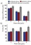
Comparison of the antibody–aptamer ensemble with antibody or aptamer alone for cell capture: (a) capture efficiency of 103 mL−1 CEM cells (target) and 106 mL−1 Ramos cells (negative control) in PBS buffer; (b) capture purity under the same conditions. Error bars represent one standard deviation (n = 3).
Fig. 4
Fig. 4
Calibration plot of cancer cell capture from whole blood and a buffer solution containing different cell concentrations (flow rate at 2.0 μL s−1); solid lines represent linear fitting. Error bars represent one standard deviation (n = 3). The inset is a representative image of captured CEM cells (DiI+, DAPI+) from whole blood; the DAPI+ cells (blue only) are non-specifically captured white blood cells.

References

    1. Maheswaran S, Sequist LV, Nagrath S, Ulkus L, Brannigan B, Collura CV, Inserra E, Diederichs S, Iafrate AJ, Bell DW, Digumarthy S, Muzikansky A, Irimia D, Settleman J, Tompkins RG, Lynch TJ, Toner M, Haber DA. N. Engl. J. Med. 2008;359:366–377. - PMC - PubMed
    1. Pantel K, Brakenhoff RH, Brandt B. Nat. Rev. Cancer. 2008;8:329–340. - PubMed
    1. Sheng W, Chen T, Tan W, Fan ZH. ACS Nano. 2013;7:7067–7076. - PMC - PubMed
    1. Zhao MX, Schiro PG, Kuo JS, Koehler KM, Sabath DE, Popov V, Feng QH, Chiu DT. Anal. Chem. 2013;85:2465–2471. - PMC - PubMed
    1. Myung JH, Gajjar KA, Saric J, Eddington DT, Hong S. Angew. Chem., Int. Ed. 2011;50:11769–11772. - PMC - PubMed

Publication types

MeSH terms