

Case Reports
doi: 10.1136/bcr-2014-204510.
An incidental talonavicular coalition in a diabetic patient: a podiatric perspective
Affiliations
- PMID: 24827664
- PMCID: PMC4024532
- DOI: 10.1136/bcr-2014-204510
Item in Clipboard
Case Reports
An incidental talonavicular coalition in a diabetic patient: a podiatric perspective
BMJ Case Rep.
.
Abstract
A tarsal coalition is a pathological union of two or more tarsal bones. The authors present an incidental finding of a unilateral talonavicular (TN) coalition that was overlooked in a 57-year-old diabetic female with signs and symptoms of peripheral neuropathy. This case highlights the clinical implications and important teaching points in recognising a TN coalition. This is particularly relevant for new, upcoming clinicians who may have never been exposed to this diagnostic rarity during clinical training.
2014 BMJ Publishing Group Ltd.
Figures

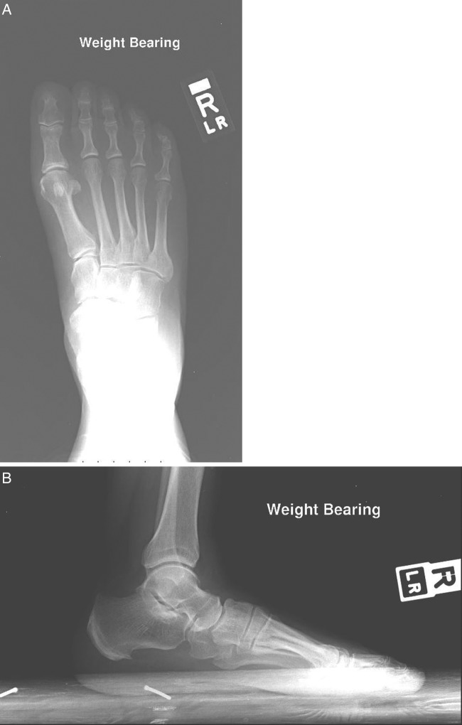
(A) (Taken in 2009): Weight bearing, anterior-posterior view of the left foot. There is no evidence of fracture or dislocation, no lytic or sclerotic lesions were noted, and there was no fusion between the talus and navicular. (B) (Taken in 2009): Weight bearing, lateral view of the left foot. The talus is not fused with the navicular.
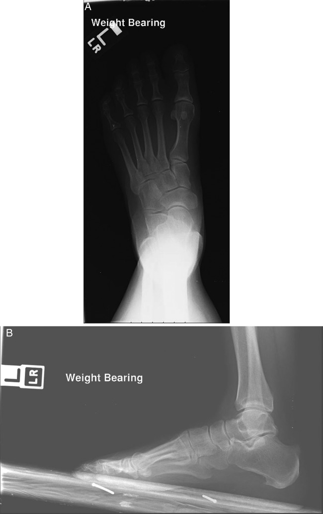
(A) (Taken in 2009): Weight bearing, anterior-posterior view of the right foot. Note the radiograph was taken at low millamperage and so the talonavicular (TN) synostosis could not be visualised. (B) (Taken in 2009): Weight bearing, lateral view of the right foot indicating a TN synostosis. A plantar calcaneal spur is also evident.
(A) (Taken in 2012): Non-weight bearing, anterior-posterior view of the right foot indicating a talonavicular (TN) synostosis. There is evidence of mild degenerative changes in the midfoot. There is no evident fracture or dislocation. (B) (Taken in 2012): Non-weight bearing, medial oblique view of the right foot indicating a TN synostosis. (C) (Taken in 2012): Non-weight bearing, lateral view of the right foot revealing a TN synostosis. A plantar calcaneal spur was also noted.
References
-
- Stormont DM, Peterson HA. The relative incidence of tarsal coalition. Clin Orthop Relat Res 1983;181:28–36 - PubMed
-
- Harris RI, Beath T. Etiology of peroneal spastic flat foot. J Bone Joint Surg Br 1948;30B:624–34 - PubMed
-
- Mosier KM, Asher M. Tarsal coalitions and peroneal spastic flat foot. A review. J Bone Joint Surg Am 1984;66:976–84 - PubMed
-
- American Diabetes Association. Standards of medical care in diabetes—2014. Diabetes Care 2014;37(Suppl 1):S14–80 - PubMed
-
- Pontious J, Hillstrom HJ, Monahan T, et al. Talonavicular coalition. Objective gait analysis. J Am Podiatr Med Assoc 1993;83:379–85 - PubMed
Publication types
MeSH terms
Supplementary concepts
LinkOut - more resources
Full Text Sources
Other Literature Sources
Medical