

A next-generation genetically attenuated Plasmodium falciparum parasite created by triple gene deletion
- PMID: 24827907
- PMCID: PMC4435496
- DOI: 10.1038/mt.2014.85
A next-generation genetically attenuated Plasmodium falciparum parasite created by triple gene deletion
Abstract
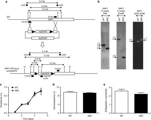
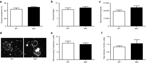
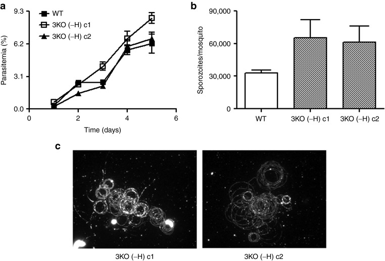
Immunization with live-attenuated Plasmodium sporozoites completely protects against malaria infection. Genetic engineering offers a versatile platform to create live-attenuated sporozoite vaccine candidates. We previously generated a genetically attenuated parasite (GAP) by deleting the P52 and P36 genes in the NF54 wild-type (WT) strain of Plasmodium falciparum (Pf p52(-)/p36(-) GAP). Preclinical assessment of p52(-)/p36(-) GAP in a humanized mouse model indicated an early and severe liver stage growth defect. However, human exposure to >200 Pf p52(-)/p36(-) GAP-infected mosquito bites in a safety trial resulted in peripheral parasitemia in one of six volunteers, revealing that this GAP was incompletely attenuated. We have now created a triple gene deleted GAP by additionally removing the SAP1 gene (Pf p52(-)/p36(-)/sap1(-) GAP) and employed flippase (FLP)/flippase recognition target (FRT) recombination for drug selectable marker cassette removal. This next-generation GAP was indistinguishable from WT parasites in blood stage and mosquito stage development. Using an improved humanized mouse model transplanted with human hepatocytes and human red blood cells, we show that despite a high-dose sporozoite challenge, Pf p52(-)/p36(-)/sap1(-) GAP did not transition to blood stage infection and appeared to be completely attenuated. Thus, clinical testing of Pf p52(-)/p36(-)/sap1(-) GAP assessing safety, immunogenicity, and efficacy against sporozoite challenge is warranted.
Figures
Similar articles
-
The Development of Whole Sporozoite Vaccines for Plasmodium falciparum Malaria.Front Immunol. 2018 Dec 11;9:2748. doi: 10.3389/fimmu.2018.02748. eCollection 2018. Front Immunol. 2018. PMID: 30619241 Free PMC article. Review.
-
First-in-human evaluation of genetically attenuated Plasmodium falciparum sporozoites administered by bite of Anopheles mosquitoes to adult volunteers.Vaccine. 2013 Oct 9;31(43):4975-83. doi: 10.1016/j.vaccine.2013.08.007. Epub 2013 Sep 8. Vaccine. 2013. PMID: 24029408 Clinical Trial.
-
Complete attenuation of genetically engineered Plasmodium falciparum sporozoites in human subjects.Sci Transl Med. 2017 Jan 4;9(371):eaad9099. doi: 10.1126/scitranslmed.aad9099. Sci Transl Med. 2017. PMID: 28053159 Clinical Trial.
-
Plasmodium yoelii sporozoites with simultaneous deletion of P52 and P36 are completely attenuated and confer sterile immunity against infection.Infect Immun. 2007 Aug;75(8):3758-68. doi: 10.1128/IAI.00225-07. Epub 2007 May 21. Infect Immun. 2007. PMID: 17517871 Free PMC article.
-
Current Challenges in the Identification of Pre-Erythrocytic Malaria Vaccine Candidate Antigens.Front Immunol. 2020 Feb 21;11:190. doi: 10.3389/fimmu.2020.00190. eCollection 2020. Front Immunol. 2020. PMID: 32153565 Free PMC article. Review.
Cited by
-
The case for a rational genome-based vaccine against malaria.Front Microbiol. 2015 Jan 22;5:741. doi: 10.3389/fmicb.2014.00741. eCollection 2014. Front Microbiol. 2015. PMID: 25657640 Free PMC article. Review.
-
Flp/FRT-mediated disruption of ptex150 and exp2 in Plasmodium falciparum sporozoites inhibits liver-stage development.Proc Natl Acad Sci U S A. 2024 Jul 9;121(28):e2403442121. doi: 10.1073/pnas.2403442121. Epub 2024 Jul 5. Proc Natl Acad Sci U S A. 2024. PMID: 38968107 Free PMC article.
-
The Development of Whole Sporozoite Vaccines for Plasmodium falciparum Malaria.Front Immunol. 2018 Dec 11;9:2748. doi: 10.3389/fimmu.2018.02748. eCollection 2018. Front Immunol. 2018. PMID: 30619241 Free PMC article. Review.
-
A Novel and Conserved Plasmodium Sporozoite Membrane Protein SPELD is Required for Maturation of Exo-erythrocytic Forms.Sci Rep. 2017 Jan 9;7:40407. doi: 10.1038/srep40407. Sci Rep. 2017. PMID: 28067322 Free PMC article.
-
Genetic approach towards a vaccine against malaria.Eur J Clin Microbiol Infect Dis. 2018 Oct;37(10):1829-1839. doi: 10.1007/s10096-018-3313-8. Epub 2018 Jun 28. Eur J Clin Microbiol Infect Dis. 2018. PMID: 29956023 Review.
References
-
- World Health Organization Malaria Control Department. (2013). Malaria report 2013 . http://www.who.int/malaria/publications/world_malaria_report_2013/en
-
- Nussenzweig RS, Vanderberg J, Most H, Orton C. Protective immunity produced by the injection of x-irradiated sporozoites of plasmodium berghei. Nature. 1967;216:160–162. - PubMed
-
- Clyde DF, Most H, McCarthy VC, Vanderberg JP. Immunization of man against sporozite-induced falciparum malaria. Am J Med Sci. 1973;266:169–177. - PubMed
-
- Clyde DF, McCarthy VC, Miller RM, Hornick RB. Specificity of protection of man immunized against sporozoite-induced falciparum malaria. Am J Med Sci. 1973;266:398–403. - PubMed
Publication types
MeSH terms
Substances
LinkOut - more resources
Full Text Sources
Other Literature Sources
Miscellaneous
