

Imaging single chiral nanoparticles in turbid media using circular-polarization optical coherence microscopy
- PMID: 24828009
- PMCID: PMC4021320
- DOI: 10.1038/srep04979
Imaging single chiral nanoparticles in turbid media using circular-polarization optical coherence microscopy
Abstract
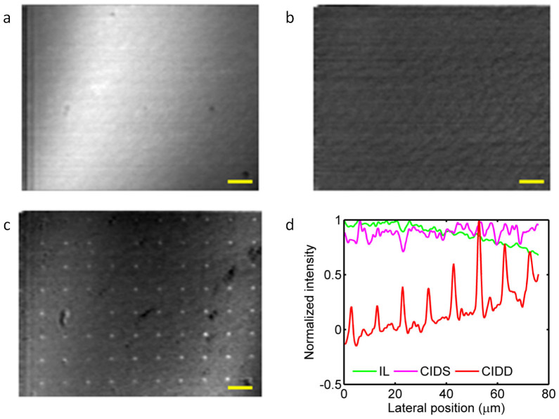
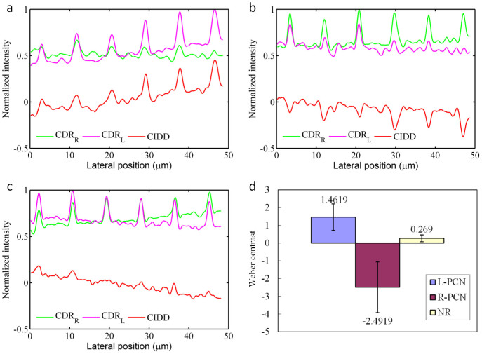
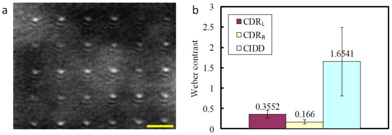
Optical coherence tomography (OCT) is a widely used structural imaging method. However, it has limited use in molecular imaging due to the lack of an effective contrast mechanism. Gold nanoparticles have been widely used as molecular probes for optical microcopy based on Surface Plasmon Resonance (SPR). Unfortunately, the SPR enhanced backscattering from nanoparticles is still relatively weak compared with the background signal from microscopic structures in biological tissues when imaged with OCT. Consequently, it is extremely challenging to perform OCT imaging of conventional nanoparticles in thick tissues with sensitivity comparable to that of fluorescence imaging. We have discovered and demonstrated a novel approach towards remarkable contrast enhancement, which is achieved by the use of a circular-polarization optical coherence microscopy system and 3-dimensional chiral nanostructures as contrast agents. By detecting the circular intensity differential depolarization (CIDD), we successfully acquired high quality images of single chiral nanoparticles underneath a 1-mm-thick tissue -mimicking phantom.
Figures
Similar articles
-
Plasmonic chiral contrast agents for optical coherence tomography: numerical study.Opt Express. 2011 Aug 1;19(16):14903-12. doi: 10.1364/OE.19.014903. Opt Express. 2011. PMID: 21934851
-
Optical coherence tomography with plasmon resonant nanorods of gold.Opt Lett. 2007 Jun 1;32(11):1438-40. doi: 10.1364/ol.32.001438. Opt Lett. 2007. PMID: 17546147
-
Tissue perfusion modelling in optical coherence tomography.Biomed Eng Online. 2017 Feb 8;16(1):27. doi: 10.1186/s12938-017-0320-4. Biomed Eng Online. 2017. PMID: 28178998 Free PMC article.
-
Advances in contrast enhancement for optical coherence tomography.Conf Proc IEEE Eng Med Biol Soc. 2006;2006:121-4. doi: 10.1109/IEMBS.2006.259366. Conf Proc IEEE Eng Med Biol Soc. 2006. PMID: 17946382 Review.
-
An overview of optical coherence tomography for ovarian tissue imaging and characterization.Wiley Interdiscip Rev Nanomed Nanobiotechnol. 2015 Jan-Feb;7(1):1-16. doi: 10.1002/wnan.1306. Epub 2014 Oct 20. Wiley Interdiscip Rev Nanomed Nanobiotechnol. 2015. PMID: 25329515 Free PMC article. Review.
Cited by
-
Aperture phase modulation with adaptive optics: a novel approach for speckle reduction and structure extraction in optical coherence tomography.Biomed Opt Express. 2019 Jan 15;10(2):552-570. doi: 10.1364/BOE.10.000552. eCollection 2019 Feb 1. Biomed Opt Express. 2019. PMID: 30800499 Free PMC article.
-
In vivo investigation of temporal effects and drug delivery induced by transdermal microneedles with optical coherence tomography.Biomed Opt Express. 2016 Apr 15;7(5):1865-76. doi: 10.1364/BOE.7.001865. eCollection 2016 May 1. Biomed Opt Express. 2016. PMID: 27231627 Free PMC article.
-
Plasmonic Nanoparticles: Advanced Researches (II).Adv Exp Med Biol. 2021;1309:161-190. doi: 10.1007/978-981-33-6158-4_7. Adv Exp Med Biol. 2021. PMID: 33782872
-
Polarization-sensitive optical projection tomography for muscle fiber imaging.Sci Rep. 2016 Jan 11;6:19241. doi: 10.1038/srep19241. Sci Rep. 2016. PMID: 26752330 Free PMC article.
-
Polarization sensitive optical coherence microscopy for brain imaging.Opt Lett. 2016 May 15;41(10):2213-6. doi: 10.1364/OL.41.002213. Opt Lett. 2016. PMID: 27176965 Free PMC article.
References
-
- Sparkes A. H., Werrett G., Stokes C. R. & Gruffyddjones T. J. Improved Sensitivity in the Diagnosis of Dermatophytosis by Fluorescence Microscopy with Calcafluor White. Vet Rec 134, 307–308 (1994). - PubMed
-
- Rolfe D. J. et al. Automated multidimensional single molecule fluorescence microscopy feature detection and tracking. Eur Biophys J Biophy 40, 1167–1186, 10.1007/s00249-011-0747-7 (2011). - PubMed
Publication types
MeSH terms
Substances
LinkOut - more resources
Full Text Sources
Other Literature Sources
