. 2014 May 22;509(7501):507-11.
doi: 10.1038/nature13324. Epub 2014 May 14.

Mfsd2a is critical for the formation and function of the blood-brain barrier

Affiliations

Mfsd2a is critical for the formation and function of the blood-brain barrier

Ayal Ben-Zvi et al. Nature. .

Abstract

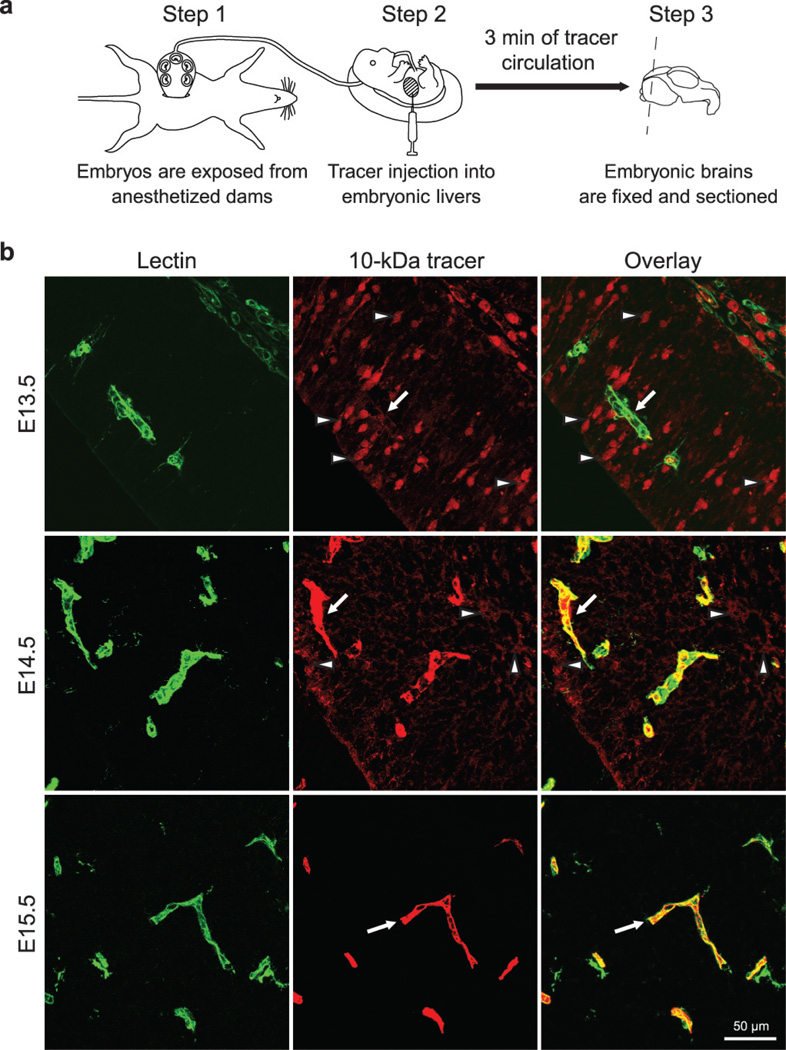
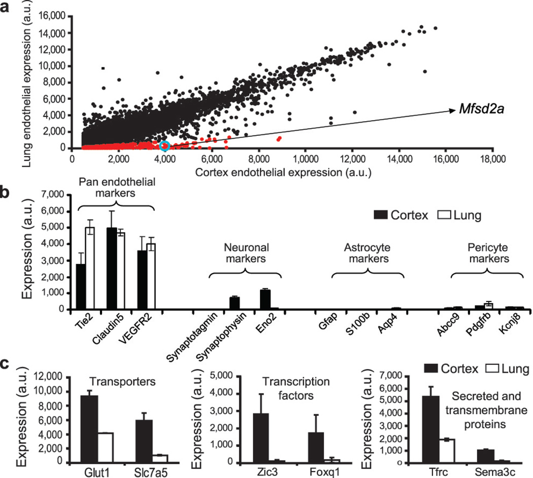
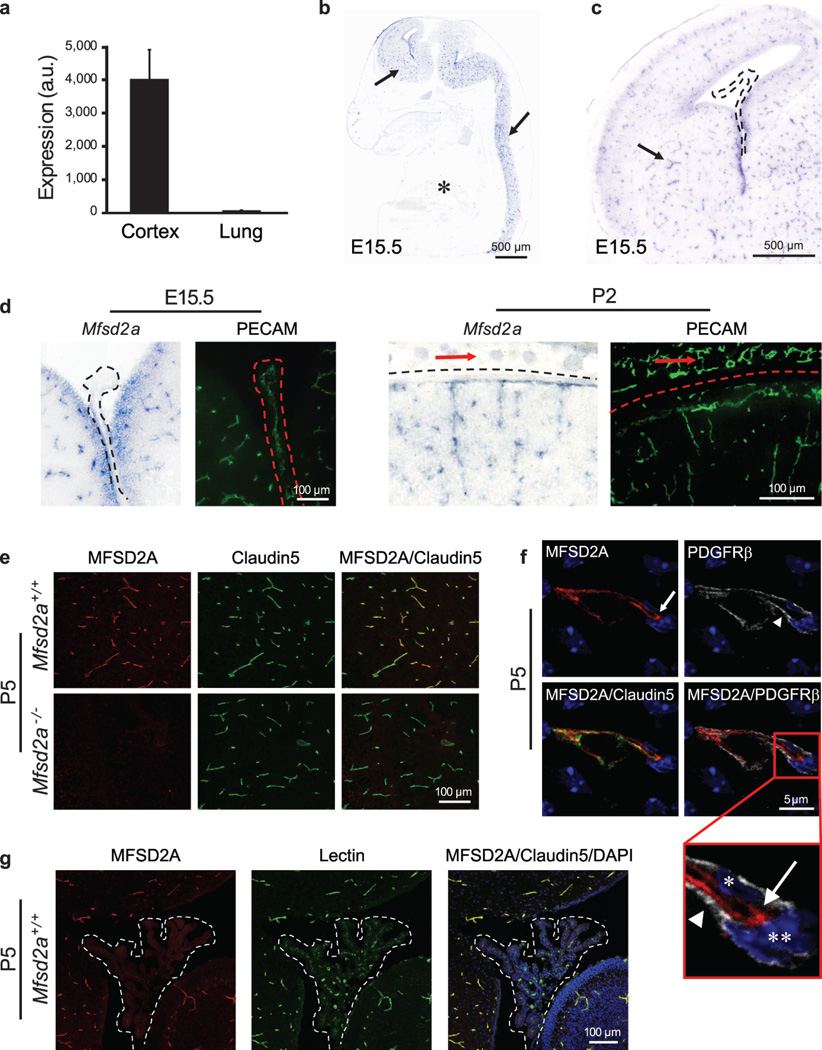
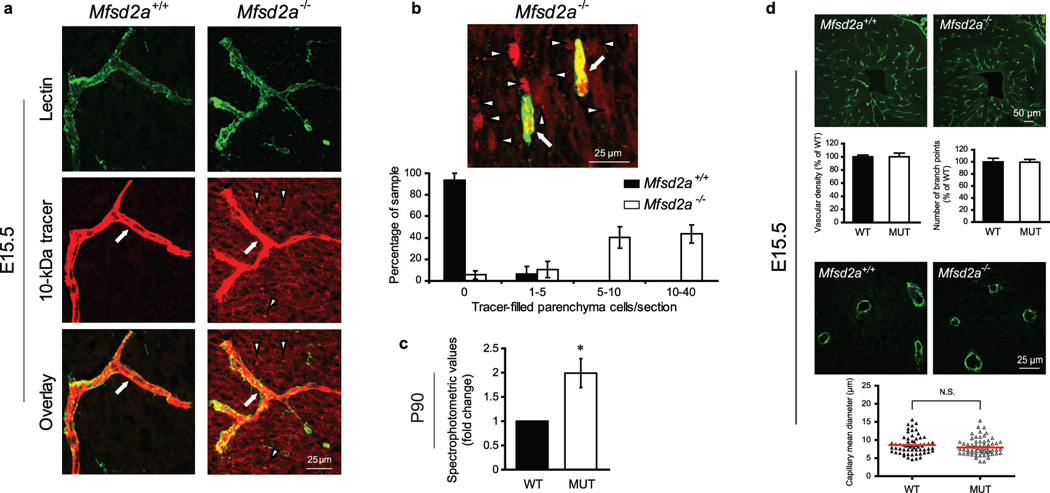
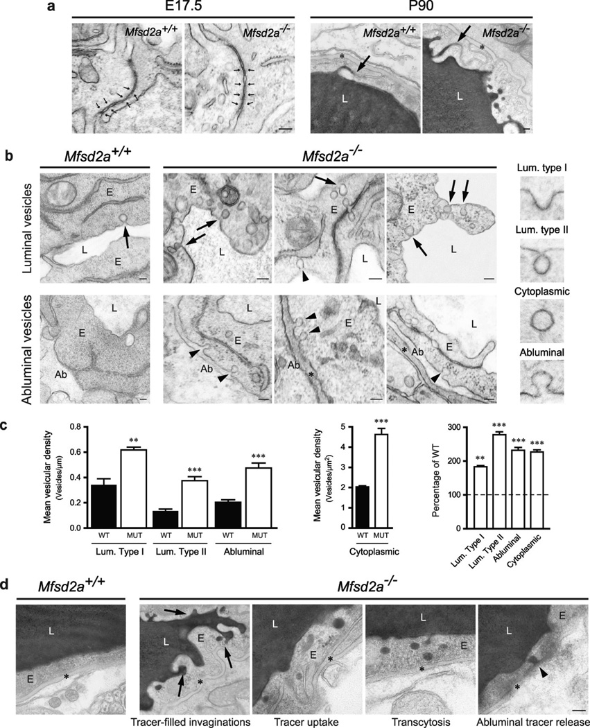
The central nervous system (CNS) requires a tightly controlled environment free of toxins and pathogens to provide the proper chemical composition for neural function. This environment is maintained by the 'blood-brain barrier' (BBB), which is composed of blood vessels whose endothelial cells display specialized tight junctions and extremely low rates of transcellular vesicular transport (transcytosis). In concert with pericytes and astrocytes, this unique brain endothelial physiological barrier seals the CNS and controls substance influx and efflux. Although BBB breakdown has recently been associated with initiation and perpetuation of various neurological disorders, an intact BBB is a major obstacle for drug delivery to the CNS. A limited understanding of the molecular mechanisms that control BBB formation has hindered our ability to manipulate the BBB in disease and therapy. Here we identify mechanisms governing the establishment of a functional BBB. First, using a novel tracer-injection method for embryos, we demonstrate spatiotemporal developmental profiles of BBB functionality and find that the mouse BBB becomes functional at embryonic day 15.5 (E15.5). We then screen for BBB-specific genes expressed during BBB formation, and find that major facilitator super family domain containing 2a (Mfsd2a) is selectively expressed in BBB-containing blood vessels in the CNS. Genetic ablation of Mfsd2a results in a leaky BBB from embryonic stages through to adulthood, but the normal patterning of vascular networks is maintained. Electron microscopy examination reveals a dramatic increase in CNS-endothelial-cell vesicular transcytosis in Mfsd2a(-/-) mice, without obvious tight-junction defects. Finally we show that Mfsd2a endothelial expression is regulated by pericytes to facilitate BBB integrity. These findings identify Mfsd2a as a key regulator of BBB function that may act by suppressing transcytosis in CNS endothelial cells. Furthermore, our findings may aid in efforts to develop therapeutic approaches for CNS drug delivery.


Figures

Figure 1
Figure 1. A novel tracer injection method reveals a temporal profile of functional BBB formation in the embryonic cortex
a, In-utero embryonic liver tracer injection method - fenestrated liver vasculature allowed rapid tracer uptake into the embryonic circulation. b, 10-kDa dextran-tracer injection revealed a temporal profile of functional cortical BBB formation. Representative images of dorsal cortical plates from injected embryos after capillary labeling with lectin (Green: lectin, red: 10-kDa tracer). Upper panel, E13.5: Tracer leaked out of capillaries and was subsequently taken up by non-vascular parenchyma cells (arrowheads), with little tracer left inside capillaries (arrow). Middle panel, E14.5: Tracer was primarily restricted to capillaries (arrow), with diffused tracer detectable in the parenchyma (arrowheads). Lower panel, E15.5: Tracer was confined to capillaries (arrow). n=6 embryos (3 litters/age).
Figure 2
Figure 2. Expression profiling identifies genes involved in BBB formation
a, Dot plot representation of Affymetrix GeneChip data showing transcriptional profile of cortical (BBB) and lung (non-BBB) endothelial cells isolated at the critical barrier-genesis period (E13.5). Dots reflect average expression of a probe in the cortex (x-axis) and lung (y-axis). Cortex expression values above 500 arbitrary expression units (a.u) are presented. Red dots indicate a 5-fold higher expression in the cortex. Mfsd2a value is circled in blue. b, Pan-endothelial markers were highly represented, whereas pericyte, astrocyte, and neuronal markers were detected at extremely low levels in both cortex and lung samples. c, Barrier-genesis specific transporters, transcription factors, and secreted and transmembrane proteins were significantly enriched in the cortical endothelial cells. All data are mean±s.d. n=4 litters (4 biological replicates).
Figure 3
Figure 3. Mfsd2a is selectively expressed in BBB-containing CNS vasculature
a, E13.5: Mfsd2a expression in cortical endothelium was ~80-fold higher than lung endothelium (microarray analysis, mean±s.d.). b-d, Specific Mfsd2a expression in BBB-containing CNS vasculature (Blue: Mfsd2a in situ hybridization, green: vessel staining (PECAM) adjacent sections). b, Mfsd2a expression in CNS vasculature (E15.5 sagittal view-brain and spinal cord, arrows), but not in non-CNS vasculature (asterisk). c, Mfsd2a expression in BBB vasculature (E15.5 cortex coronal view, e.g. striatum, arrow), but not in non-BBB CNS vasculature (choroid plexus, dotted line). d, High magnification coronal view of Mfsd2a expression in BBB-containing CNS vasculature but not in vasculature of the choroid plexus (left, dotted line), or outer meninges/skin (right, red arrows). e-g, Immunohistochemical staining of MFSD2A protein shows specific expression in CNS endothelial cells (Red: MFSD2A; green: Claudin5 or Lectin (endothelium); blue: DAPI (nuclei); gray: PDGFRβ (pericytes)). e, MFSD2A expression in the brain vasculature of wild-type mice (upper panel), but not of Mfsd2a−/− mice (lower panel). f, MFSD2A expression only in Claudin5-positive endothelial cells (arrow, endothelial nucleus–asterisk) but not in adjacent pericytes (arrow head, pericyte nucleus–double asterisk). g, Lack of MFSD2A expression in choroid plexus vasculature (fourth ventricle coronal view-dotted line), as opposed to the prominent MFSD2A expression in cerebellar vasculature. n=3 embryos (3 litters/age).
Figure 4
Figure 4. Mfsd2a is required for the establishment of a functional BBB but not for CNS vascular patterning in vivo
a,b, 10-kDa dextran-tracer injections at E15.5 revealed a defective BBB in mice lacking Mfsd2a. a, The tracer was confined to the capillaries (arrow) in wild-type littermates, whereas Mfsd2a−/− embryos showed large amounts of tracer leakage in the brain parenchyma (arrowheads). b, Capillaries (arrows) surrounded by tracer-filled brain parenchyma cells (arrowheads) in Mfsd2a−/− cortex. Quantification of tracer-filled parenchyma cells in control versus Mfsd2a−/− cortical plates (lower panel n=7 embryos/genotype). c, Spectrophotometric quantification of 10-kDa dextran-tracer from cortical extracts, 16hr post i.v. injection, indicating that BBB leakiness in Mfsd2a−/− mice persists into adulthood (n=3 mice/genotype). d, Mfsd2a−/− mice exhibit normal vascular patterning. No abnormalities were found in cortical vascular density, branching and capillary diameter (E15.5, green: PECAM). Quantification of wild-type and Mfsd2a−/− samples (n=4 embryos/per genotype, (P>0.5)). All data are mean±s.e.m.
Figure 5
Figure 5. Mfsd2a is required specifically to suppress transcytosis in brain endothelium to maintain BBB integrity
Electron-microscopic examination of BBB integrity. a, Embryonic Mfsd2a−/− endothelium showed no overt tight-junction ultrastructural defect (normal “kissing points”, small arrows, left). Vessel lumen in HRP-injected adult mice was filled with electron-dense DAB reaction (black) that diffused into intercellular clefts but stopped sharply at the junction without parenchymal leakage (arrows, right). b, Increased vesicular activity in embryonic Mfsd2a−/− endothelium (E17.5). Left, wild-type endothelium displayed very few vesicles (arrow). Right, Mfsd2a−/− endothelium contained many vesicles of various types: luminal-(arrows), abluminal-(arrowheads) membrane-connected and cytoplasmic vesicles. c, Vesicular density quantification (as illustrated in b, see also Fig.S7a). d, Increased transcytosis was evident in HRP-injected adult Mfsd2a−/− mice (P90). In wildtype littermates (left) HRP activity was confined to the lumen with no HRP-filled vesicles. Many HRP-filled vesicles found in Mfsd2a−/− endothelial cells (right, see quantification Fig.S7b). Luminal invaginations (dye uptake, arrows) and release to the basement membrane (abluminal side (*)). Ab: abluminal, E: endothelium, L: lumen. Scale bars: a,b: 100 nm; c, 200 nm. All data are mean±s.e.m.

Comment in

References

    1. Saunders NR, Liddelow SA, Dziegielewska KM. Barrier mechanisms in the developing brain. Front Pharmacol. 2012;3:46. - PMC - PubMed
    1. Reese TS, Karnovsky MJ. Fine structural localization of a blood-brain barrier to exogenous peroxidase. J Cell Biol. 1967;34:207–217. - PMC - PubMed
    1. Siegenthaler JA, Sohet F, Daneman R. 'Sealing off the CNS': cellular and molecular regulation of blood-brain barriergenesis. Curr Opin Neurobiol. 2013;13:001256. - PMC - PubMed
    1. Daneman R, Zhou L, Kebede AA, Barres BA. Pericytes are required for blood– brain barrier integrity during embryogenesis. Nature. 2010;468:562–566. - PMC - PubMed
    1. Armulik A, et al. Pericytes regulate the blood–brain barrier. Nature. 2010;468:557–561. - PubMed

Publication types

MeSH terms

Associated data