

Potential for adult-based epidemiological studies to characterize overall cancer risks associated with a lifetime of CT scans
- PMID: 24828111
- PMCID: PMC4157352
- DOI: 10.1667/RR13622.1
Potential for adult-based epidemiological studies to characterize overall cancer risks associated with a lifetime of CT scans
Abstract
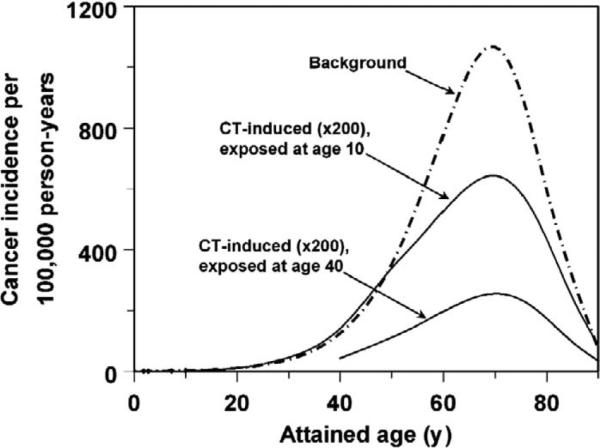
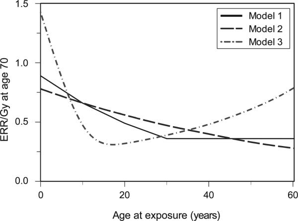
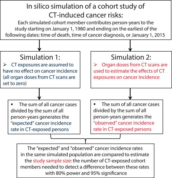
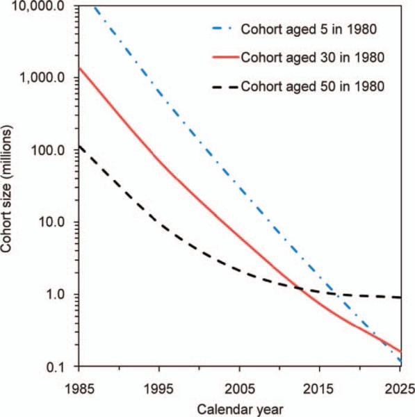
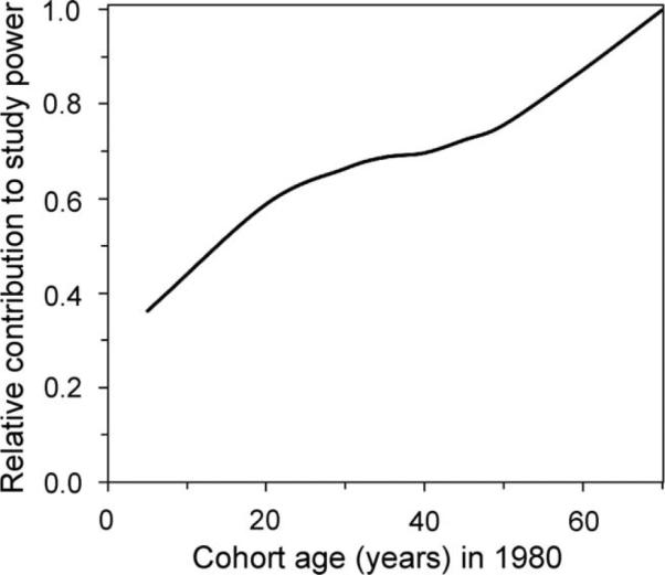
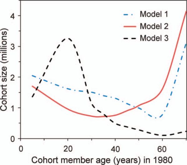
Recent epidemiological studies have suggested that radiation exposure from pediatric CT scanning is associated with small excess cancer risks. However, the majority of CT scans are performed on adults, and most radiation-induced cancers appear during middle or old age, in the same age range as background cancers. Consequently, a logical next step is to investigate the effects of CT scanning in adulthood on lifetime cancer risks by conducting adult-based, appropriately designed epidemiological studies. Here we estimate the sample size required for such studies to detect CT-associated risks. This was achieved by incorporating different age-, sex-, time- and cancer type-dependent models of radiation carcinogenesis into an in silico simulation of a population-based cohort study. This approach simulated individual histories of chest and abdominal CT exposures, deaths and cancer diagnoses. The resultant sample sizes suggest that epidemiological studies of realistically sized cohorts can detect excess lifetime cancer risks from adult CT exposures. For example, retrospective analysis of CT exposure and cancer incidence data from a population-based cohort of 0.4 to 1.3 million (depending on the carcinogenic model) CT-exposed UK adults, aged 25-65 in 1980 and followed until 2015, provides 80% power for detecting cancer risks from chest and abdominal CT scans.
Figures
Similar articles
-
Radiation exposure from CT scans in childhood and subsequent risk of leukaemia and brain tumours: a retrospective cohort study.Lancet. 2012 Aug 4;380(9840):499-505. doi: 10.1016/S0140-6736(12)60815-0. Epub 2012 Jun 7. Lancet. 2012. PMID: 22681860 Free PMC article.
-
Projected Lifetime Cancer Risks From Current Computed Tomography Imaging.JAMA Intern Med. 2025 Jun 1;185(6):710-719. doi: 10.1001/jamainternmed.2025.0505. JAMA Intern Med. 2025. PMID: 40227719 Free PMC article.
-
The use of computed tomography in pediatrics and the associated radiation exposure and estimated cancer risk.JAMA Pediatr. 2013 Aug 1;167(8):700-7. doi: 10.1001/jamapediatrics.2013.311. JAMA Pediatr. 2013. PMID: 23754213 Free PMC article.
-
[Exposure to CT scans in childhood and long-term cancer risk: A review of epidemiological studies].Bull Cancer. 2016 Feb;103(2):190-8. doi: 10.1016/j.bulcan.2015.11.003. Epub 2016 Jan 15. Bull Cancer. 2016. PMID: 26782078 Review. French.
-
Cancer risks from diagnostic radiology.Br J Radiol. 2008 May;81(965):362-78. doi: 10.1259/bjr/01948454. Br J Radiol. 2008. PMID: 18440940 Review.
Cited by
-
Patient understanding of radiation risk from medical computed tomography-A comparison of Hispanic vs. non-Hispanic emergency department populations.PeerJ. 2015 May 7;3:e937. doi: 10.7717/peerj.937. eCollection 2015. PeerJ. 2015. PMID: 26019999 Free PMC article.
-
Patient-Specific Organ and Effective Dose Estimates in Adult Oncologic CT.AJR Am J Roentgenol. 2020 Apr;214(4):738-746. doi: 10.2214/AJR.19.21197. Epub 2019 Aug 15. AJR Am J Roentgenol. 2020. PMID: 31414882 Free PMC article.
-
A Pilot Study of Prediction of Creatinine Clearance by Ellipsoid Volumetry of Kidney Using Noncontrast Computed Tomography.JMA J. 2019 Mar 4;2(1):60-66. doi: 10.31662/jmaj.2018-0021. Epub 2019 Feb 20. JMA J. 2019. PMID: 33681514 Free PMC article.
-
Is there Unmeasured Indication Bias in Radiation-Related Cancer Risk Estimates from Studies of Computed Tomography?Radiat Res. 2018 Feb;189(2):128-135. doi: 10.1667/RR14807.1. Epub 2017 Dec 5. Radiat Res. 2018. PMID: 29206598 Free PMC article.
References
-
- Brenner DJ, Hall EJ. Computed tomography–an increasing source of radiation exposure. N Engl J Med. 2007;357:2277–84. - PubMed
-
- Hricak H, Brenner DJ, Adelstein SJ, Frush DP, Hall EJ, Howell RW, et al. Managing radiation use in medical imaging: a multifaceted challenge. Radiology. 2011;258:889–905. - PubMed
-
- Sodickson A, Baeyens PF, Andriole KP, Prevedello LM, Nawfel RD, Hanson R, et al. Recurrent CT, cumulative radiation exposure, and associated radiation-induced cancer risks from CT of adults. Radiology. 2009;251:175–84. - PubMed
-
- Hansen J, Jurik AG. Analysis of current practice of ct examinations. Acta Oncol. 2009;48:295–301. - PubMed
-
- Health risks from exposure to low levels of ionizing radiation: BEIR VII phase 2. National Academies Press; National Research Council (U.S.); Washington, D.C.: 2006. Committee to Assess Health Risks from Exposure to Low Level of Ionizing Radiation. - PubMed
Publication types
MeSH terms
Grants and funding
LinkOut - more resources
Full Text Sources
Other Literature Sources
Medical
