Cytokine and nitric oxide levels in patients with sepsis--temporal evolvement and relation to platelet mitochondrial respiratory function
- PMID: 24828117
- PMCID: PMC4020920
- DOI: 10.1371/journal.pone.0097673
Cytokine and nitric oxide levels in patients with sepsis--temporal evolvement and relation to platelet mitochondrial respiratory function
Erratum in
-
Cytokine and nitric oxide levels in patients with sepsis--temporal evolvement and relation to platelet mitochondrial respiratory function.PLoS One. 2014 Jul 22;9(7):e103756. doi: 10.1371/journal.pone.0103756. eCollection 2014. PLoS One. 2014. PMID: 25051116 Free PMC article.
Abstract
Background: The levels of nitric oxide (NO) and various cytokines are known to be increased during sepsis. These signaling molecules could potentially act as regulators and underlie the enhancement of mitochondrial function described in the later phase of sepsis. Therefore, we investigated the correlation between observed changes in platelet mitochondrial respiration and a set of pro- and anti-inflammatory cytokines as well as NO plasma levels in patients with sepsis.
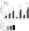
Methods and results: Platelet mitochondrial respiration and levels of TNFα, MCP-1 (monocyte chemotactic protein-1), INFγ (interferon-γ), IL-1β, IL-4, IL-5, IL-6, IL-8, IL-10 and IL-17 and NO were analyzed in 38 patients with severe sepsis or septic shock at three time points during one week following admission to the ICU. Citrate synthase, mitochondrial DNA and cytochrome c were measured as markers of cellular mitochondrial content. All mitochondrial respiratory states increased over the week analyzed (p<0.001). IL-8 levels correlated with maximal mitochondrial respiration on day 6-7 (p = 0.02, r2 = 0.22) and was also higher in non-survivors compared to survivors on day 3-4 and day 6-7 (p = 0.03 respectively). Neither NO nor any of the other cytokines measured correlated with respiration or mortality. Cytochrome c levels were decreased at day 1-2 by 24±5% (p = 0.03) and returned towards values of the controls at the last two time points. Citrate synthase activity and mitochondrial DNA levels were similar to controls and remained constant throughout the week.
Conclusions: Out of ten analyzed cytokines and nitric oxide, IL-8 correlated with the observed increase in mitochondrial respiration. This suggests that cytokines as well as NO do not play a prominent role in the regulation of platelet mitochondrial respiration in sepsis. Further, the respiratory increase was not accompanied by an increase in markers of mitochondrial content, suggesting a possible role for post-translational enhancement of mitochondrial respiration rather than augmented mitochondrial mass.
Conflict of interest statement
Figures


References
-
- Vincent JL, Nelson DR, Williams MD (2011) Is worsening multiple organ failure the cause of death in patients with severe sepsis? Crit Care Med 39: 1050–1055. - PubMed
-
- Fry DE (2012) Sepsis, systemic inflammatory response, and multiple organ dysfunction: the mystery continues. Am Surg 78: 1–8. - PubMed
-
- Brealey D, Singer M (2003) Mitochondrial Dysfunction in Sepsis. Curr Infect Dis Rep 5: 365–371. - PubMed
Publication types
MeSH terms
Substances
LinkOut - more resources
Full Text Sources
Other Literature Sources
Research Materials
Miscellaneous

