Review
. 2014 May 13;12(5):2877-912.
doi: 10.3390/md12052877.

Evolving marine biomimetics for regenerative dentistry

Affiliations
Review

Evolving marine biomimetics for regenerative dentistry

David W Green et al. Mar Drugs. .

Abstract

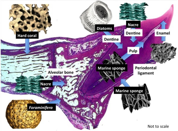
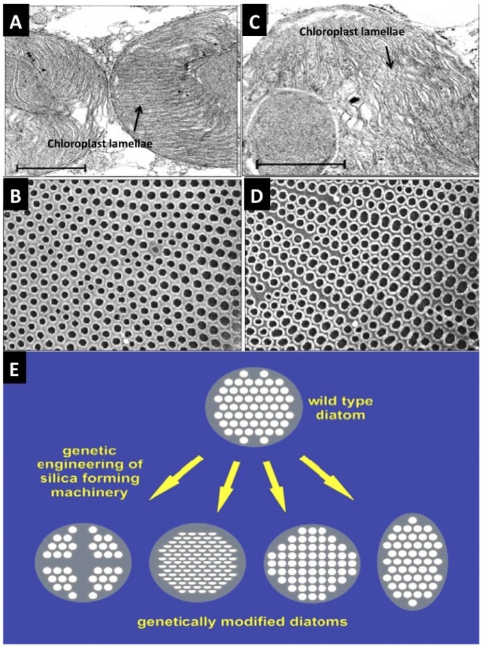
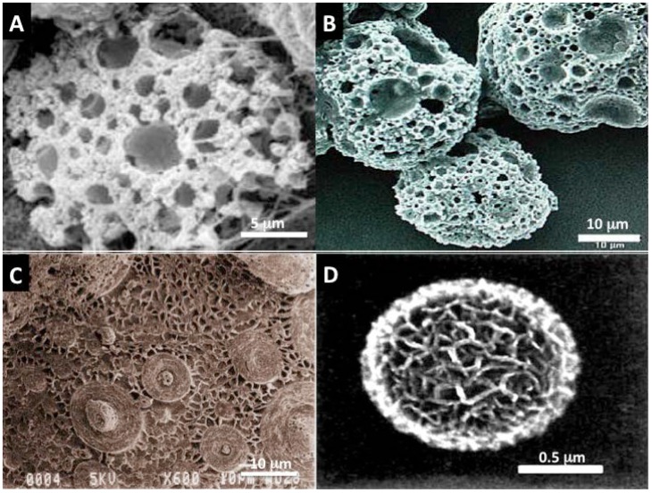
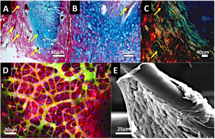
New products that help make human tissue and organ regeneration more effective are in high demand and include materials, structures and substrates that drive cell-to-tissue transformations, orchestrate anatomical assembly and tissue integration with biology. Marine organisms are exemplary bioresources that have extensive possibilities in supporting and facilitating development of human tissue substitutes. Such organisms represent a deep and diverse reserve of materials, substrates and structures that can facilitate tissue reconstruction within lab-based cultures. The reason is that they possess sophisticated structures, architectures and biomaterial designs that are still difficult to replicate using synthetic processes, so far. These products offer tantalizing pre-made options that are versatile, adaptable and have many functions for current tissue engineers seeking fresh solutions to the deficiencies in existing dental biomaterials, which lack the intrinsic elements of biofunctioning, structural and mechanical design to regenerate anatomically correct dental tissues both in the culture dish and in vivo.


Figures

Figure 1
Figure 1
Structural products harnessed directly from marine life. In this show a small selection of marine biostructures with promising properties to rebuild the main individual tooth tissues that are affected by disease: bone, dentine, pulp and periodontal ligament. A further look into marine natural history will offer more structures destined for transplantation into the tooth structure itself. These marine derived structures are required to support dental cells and tissues in their natural position and create structural, crystalline and mineral ion clustered microenvironments for developing mineralized and connective tissues with properties that ensure seamless fusion takes place within existing tooth structures. Tissue boosting molecules such as, growth factor proteins can be stably adsorbed into the marine substitutes and also be entrapped within the mesopores and nanopores. Various routes for appropriating the structures can be employed that increase the number and type of biological roles they have. We can add more roles than the original structure to jump start regeneration.
Figure 2
Figure 2
Ideas for re-design of the structure of microshells via genetic and metabolic routes. The architectural features of Coscinodiscus wailesii species of Diatom frustules can be changed via modulation of the chemical and biochemical composition of the growing environment [77]. (A) TEM cross-section through chloroplasts in Diatom cell to show the normal ultrastructure of stacked lamellae. This exact arrangement of organelles molds the areas where silica is deposited and hardened to produce a regular externalized pore structure (B); (C,D) Added Nickel sulfate-in “sub-lethal quantities”-in seawater distorts the shape and packing arrangement of lamellae, which in turn affects silica pattern formation at the cell surface. The result of this is to double the pore size and increases the packing density of the pores. This property is something we want to exploit. Gene modulation of Thalassiosira pseudonana has led to the directed evolution of differently structured Diatoms with stark variations in pore size and patterns of pores [78]. This can be a basis for adapting inorganic structures and architectures to desired formats.
Figure 3
Figure 3
Simple inorganic “designoid” objects [76] in nature recreated in the beaker using biomimetic template mediated materials chemistry (A,B) Mesoporous vaterite microsponges produced via mineralization at the interface of a SDS stabilized water microdroplet in oil; Such structures have also been manufactured using plant based surfactants and oils. The stable emulsion is reminiscent of a “viniagrette“ [83,84]; (C) Inorganic surface with multiple patterns that are strongly reminiscent of Diatoms and Radiolaria shells, created by heat treated mesolamellar aluminophosphate vesicles [80]; (D) A single mesoporous sphere of aragonite polymorph of calcium carbonate. The principle behind its formation is analogous to the arrangement of areolar vesicles in diatom mineralization. A thin soapy foam is created from an oil and water mixture. This serves as a template for calcium carbonate mineralization [80]. Using some basic construction principles learnt from nature such as: make vesicles and pack them together like foams and use them as boundaries for mineralization to build on, it is then possible to generate some of the structures and architectural motifs seen amongst Coccolithophores and Foraminifera shells [80,81,83]. These microsponges by virtue of their shape and structure have been successful in drug and gene delivery experiments and in 3-dimensional co-culture with human bone marrow stromal cells.
Figure 4
Figure 4
Collagenous Marine sponge comprises of a fibrous framework of bonded fibers and this could be an ideal substitute for a periodontal ligament and bone tissue (AC). The advantage of selected marine sponge skeletons over many synthetic fiber networks and electrospun constructs is that they are fully fused together. In (A) we see rapid colonisation, proliferation of periodontal ligament stem cells on a collagenous marine sponge within 48 h of dynamic seeding (×200). The PDL stem cells were centrifuged into the marine sponge framework at 1200 rpm for minutes. The fibers provide surfaces for periodontal ligament stem cells to develop a stretched morphology, which is characteristic of their home environment and they span large spaces existing between sponge fibers in all directions. An early form of bone and cartilage tissue is developed in vivo within a collagenous marine sponge skeleton from seeded embryonic stem cells (B,C). The histoarchitecture is early in origin and the organisation is characteristic of osteoid type of bone and cartilage. The bone that was formed showed highly organized matrix when viewed under polarized light, hinting at the possibility of highly organized mineralization in progress (Yellow arrows point to sponge fiber) (4AC Kindly reprinted from Biomedical Materials [38]); (D) Confocal fluorescence image of osteoblast cell sheets attached and suspended in marine sponge framework at 14 days. The top panel showing cell distribution at high power and the lower panel showing coverage across the whole seeded marine sponge skeleton (red = F-actin staining; green = autofluorescence of sponge fiber; blue = nucleus staining); (E) SEM image of osteoblast cell aggregation on Hippospongia fiber; (Figure D was reproduced with kind permission from Ivyspring International Publisher [54] and Figure E reproduced with kind permission from Wiley-VCH [129]).
Figure 5
Figure 5
A Diatom shell structure that can deliver incorporated antibiotic. (A) A low power SEM of one species of Diatom shell (cylindrical Aulacoseira sp.) from diatomaceous earth; (B) High power SEM of a Diatom shell to show the nanopores incorporated into the microstructure of discoid Coscinodiscus wailesii; (C) A cumulative release profile for shell incorporated Gentamicin antibiotic lasting up to 14 days before all of the product has been off-loaded. This demonstrates that Diatom shells possess a potential for controlled drug delivery (C kindly reprinted with permission from Future Medicine) [132].
Figure 6
Figure 6
Foraminifera spheres are macroscopic shells from marine sediments that grow a highly intricate set of pore structures for the task of passive filtration where food particles are sucked in through channels and by design selected into sizes for the cell inside; (A) The natural structure of interconnected pores and channels has shown a usefulness in tissue engineering for capturing and slowly releasing bone building drugs and supporting proliferation and expansion of stem cells around the outer surface in 3-dimensions (Image generated at the Microstructural Analysis Unit UTS, Sydney, Australia); (B) Close-in cross-sectional view of the macrosphere to show the regular and highly ordered distribution of pores inside the sphere. This image was generated by micro-Tomography (micro-CT at the Australian Centre for Microscopy and Microanalysis); (C) A look at the functional release of Gentamicin antibiotic from macrospheres on the eradication of S. aureus bacteria. At 30 min all bacteria cultured with Gentamicin laden Foraminifera macrospheres were exterminated; (D) An SEM image of a single macrosphere coated with rat adipocyte derived stem cells (ADSC) 7 days after seeding (C Reproduced with kind permission from Future Medicine and amended to improve clarity in reproduction [156] and D Reproduced with kind permission from Anne-Liebert [79]).
Figure 7
Figure 7
(A) Whole nacre incisor replacements found in the lower jaw of an Ancient Mayan individual. Excellent bone fusion is shown by X-ray imaging in some of the nacre implants in panels, b and c; (B) A high power (×600) image of a section at the interface between a nacre piece and metabolically active mesenchymal cells mobilizing into an osteoprogenitor layer. The junction is free of immune cells are present and fibrous tissue [162]; (C) Stained histological sections from human patient biopsy showing the integration between bone and nacre coupled with new bone formation; (D) A high power view of new bone at the nacre surface showing particularly the stained osteoid borders. Also generated are two textures of mature human bone: woven and lamellar; (Plate A, reprinted with kind permission from Bulletin of the History of Dentistry, Copyright (1972) [160]; Plate B reproduced with kind permission of Elsevier [162]. Plates C,D Reproduced with kind permission from Macmillan Publishing Group [115]).
Figure 8
Figure 8
The effectiveness of nacre WSM in bone formation, remodeling potential and osteoporosis treatment; (A,B) Nacre powder and WSM extract induce osteoblast mineralization via consequential molecular signaling pathways e.g., Fos-1, p-ERK, p-JUNK and up-regulate biomineralisation promoting genes BMP-2, cbfa-1 etc. Osteoclast gene activities are suppressed (C). Nacre products also prevent osteoporotic bone in OVX mice (D) (A–D reproduced with kind permission from Elsevier) [165].
Figure 9
Figure 9
Live animal bone repair using coral skeletons (untransformed) laden with mesenchymal stem cells. (A) A comparison of host bone reactions in histological stained section to a segmental defect (control), a segmental defect plugged with coral skeleton (Coral alone), plugged with coral skeleton and FBM and Coral skeleton filled with MSCs. In the group containing coral with MSCs the defect is bridged and repaired in full thickness; (B) X-ray slides comparing the mineralized bone replacement between coral skeleton with FBM and coral skeleton laden with MSCs. Only in the corals with MSCs do we get new mature bone replacement (Reprinted with kind permission from MacMillan Publishing Group) [175].
Figure 10
Figure 10
In building living tooth replacement tissues, addressing issues of mechanical design is paramount. The most potent lesson to learn from nature is the design of smart interfaces. In the right panel is show changes in the strength of compressive force across the whole length of a single tooth. Interfaces between each tissue with different individual structure and composition deflect the force from above without leading to catastrophic failure of the materials involved (Reproduced with kind permission from Elsevier with new labels added) [179].
Figure 11
Figure 11
Major application areas for marine biostructures in modern dentistry that cover engineering of entire dental tissues and tissue complexes, disintegration of biofilms, repeat mineralisation of the tooth surface-enamel and shallow dentine defects- and bone substitutes.

Similar articles

Cited by

References

    1. Williams D.F. On the nature of biomaterials. Biomaterials. 2009;30:5897–5909. doi: 10.1016/j.biomaterials.2009.07.027. - DOI - PubMed
    1. Huebsch N., Mooney D.J. Inspiration and application in the evolution of biomaterials. Nature. 2009;462:426–432. doi: 10.1038/nature08601. - DOI - PMC - PubMed
    1. Place E.S., Evans N.D., Stevens M.M. Complexity in biomaterials for tissue engineering. Nat. Mater. 2009;8:457–470. doi: 10.1038/nmat2441. - DOI - PubMed
    1. Fadeeva E., Deiwick A., Chichkov B., Schlie-Wolter S. Impact of laser-structured biomaterial interfaces on guided cell responses. Interface Focus. 2014;4:20130048. doi: 10.1098/rsfs.2013.0048. - DOI - PMC - PubMed
    1. Vadgama P. Surfaces and Interfaces for Biomaterials. CRC Press, Woodhead Pubulisher Ltd.; Boca Raton, Cambridge, England: 2005. p. 802.

Publication types

LinkOut - more resources