

Mathematical modelling of spatio-temporal cell dynamics in colonic crypts following irradiation
- PMID: 24828339
- PMCID: PMC6496481
- DOI: 10.1111/cpr.12110
Mathematical modelling of spatio-temporal cell dynamics in colonic crypts following irradiation
Abstract
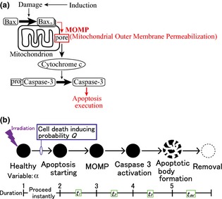
Objectives: Modelling the apoptotic process is essential for simulating and understanding tumour growth, as most tumour tissues carry mutations in apoptotic signalling pathways. Thus here, we have aimed to construct a mathematical model of colonic crypts that explicitly incorporates the apoptotic mechanism.
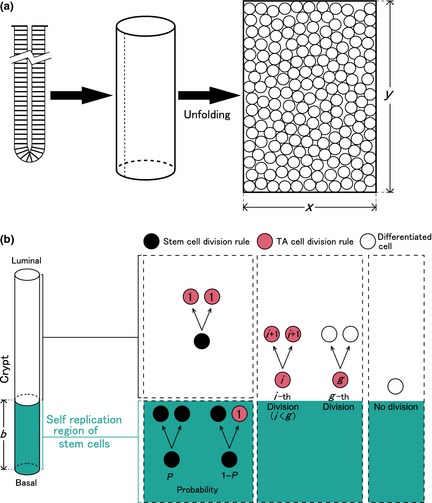
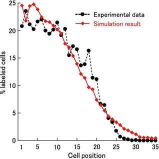
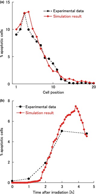
Methods: A murine colonic crypt was described as being a two-dimensional rectangular surface model. In this system, three types of cells with different proliferating and differentiating potentials migrate. Apoptosis was described as a process activated by irradiation that progresses in a stepwise manner. Parameter values in the model were determined to be consistent with experimental data for changes in the apoptotic cell ratio within murine transverse colonic crypts following irradiation.
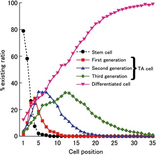
Results: First, we constructed a model reproducing cell proliferation dynamics in normal murine colonic crypts; next, we applied the apoptotic mechanism to this model. As a result, we succeeded in simultaneous reproduction of both spatial and temporal changes in distribution of apoptotic cells in murine colonic crypts by determining parameter values in numerical simulations. Through this adjustment process, we were able to predict that stem cells and transit amplifying (TA) cells in each generation must react distinctly from each other, to apoptosis-inducing stimuli.
Conclusions: We constructed a mathematical model with which we could quantitatively describe cell proliferative and apoptotic dynamics in a murine colonic crypt. Using this model, we were able to make novel predictions that sensitivity to apoptosis-inducing stimuli is dependent on cell type.
© 2014 John Wiley & Sons Ltd.
Figures
Similar articles
-
Regional differences in stem and transit cell proliferation and apoptosis in the terminal ileum and colon of mice after 12 Gy.Int J Radiat Oncol Biol Phys. 2012 Mar 1;82(3):e521-8. doi: 10.1016/j.ijrobp.2011.07.015. Epub 2011 Dec 21. Int J Radiat Oncol Biol Phys. 2012. PMID: 22196132
-
Modeling of stem cell dynamics in human colonic crypts in silico.J Gastroenterol. 2014 Feb;49(2):263-9. doi: 10.1007/s00535-013-0887-x. J Gastroenterol. 2014. PMID: 24077809
-
A calibrated agent-based computer model of stochastic cell dynamics in normal human colon crypts useful for in silico experiments.Theor Biol Med Model. 2013 Nov 18;10:66. doi: 10.1186/1742-4682-10-66. Theor Biol Med Model. 2013. PMID: 24245614 Free PMC article.
-
The evolution of metapopulation dynamics and the number of stem cells in intestinal crypts and other tissue structures in multicellular bodies.Evol Appl. 2020 Aug 12;13(7):1771-1783. doi: 10.1111/eva.13069. eCollection 2020 Aug. Evol Appl. 2020. PMID: 32821281 Free PMC article. Review.
-
Apoptosis, anoikis and their relevance to the pathobiology of colon cancer.Pathol Int. 2000 Apr;50(4):273-9. doi: 10.1046/j.1440-1827.2000.01047.x. Pathol Int. 2000. PMID: 10849312 Review.
Cited by
-
Multiplicity of Mathematical Modeling Strategies to Search for Molecular and Cellular Insights into Bacteria Lung Infection.Front Physiol. 2017 Aug 30;8:645. doi: 10.3389/fphys.2017.00645. eCollection 2017. Front Physiol. 2017. PMID: 28912729 Free PMC article. Review.
-
Optimization of Dose Schedules for Chemotherapy of Early Colon Cancer Determined by High-Performance Computer Simulations.Cancer Inform. 2019 Jan 10;18:1176935118822804. doi: 10.1177/1176935118822804. eCollection 2019. Cancer Inform. 2019. PMID: 30675100 Free PMC article.
References
-
- Meinzer HP, Sandblad B (1985) A simulation model for studies of intestine cell dynamics. Comput. Methods Programs Biomed. 21, 89–98. - PubMed
-
- Sato T, Vries RG, Snippert HJ, van de Wetering M, Barker N, Stange DE et al (2009) Single Lgr5 stem cells build crypt‐villus structures in vitro without a mesenchymal niche. Nature 459, 262–265. - PubMed
-
- Loeffler M, Stein R, Wichmann HE, Potten CS, Kaur P, Chwalinski S (1986) Intestinal cell proliferation. I. A comprehensive model of steady‐state proliferation in the crypt. Cell Tissue Kinet. 19, 627–645. - PubMed
Publication types
MeSH terms
LinkOut - more resources
Full Text Sources
Other Literature Sources
Research Materials
