. 2014 May 14;9(5):e97768.
doi: 10.1371/journal.pone.0097768. eCollection 2014.

Distribution of peripheral PrP(Sc) in sheep with naturally acquired scrapie

Affiliations

Distribution of peripheral PrP(Sc) in sheep with naturally acquired scrapie

María Carmen Garza et al. PLoS One. .

Abstract

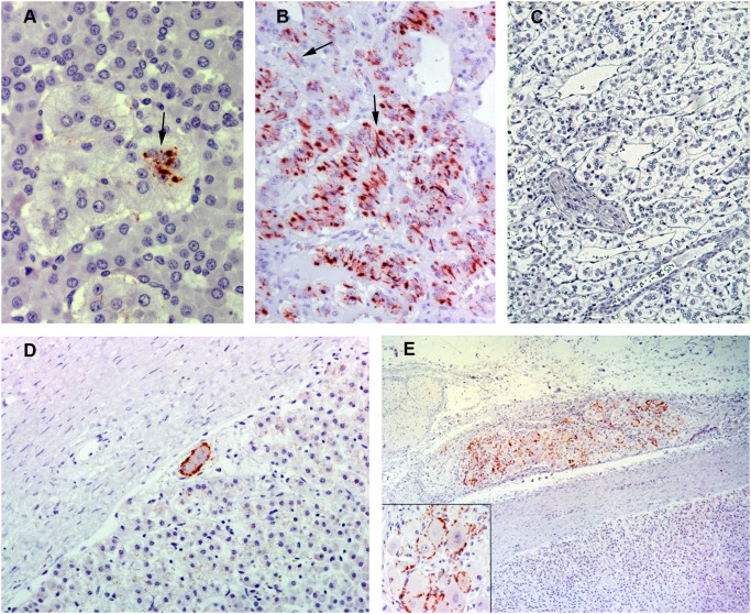
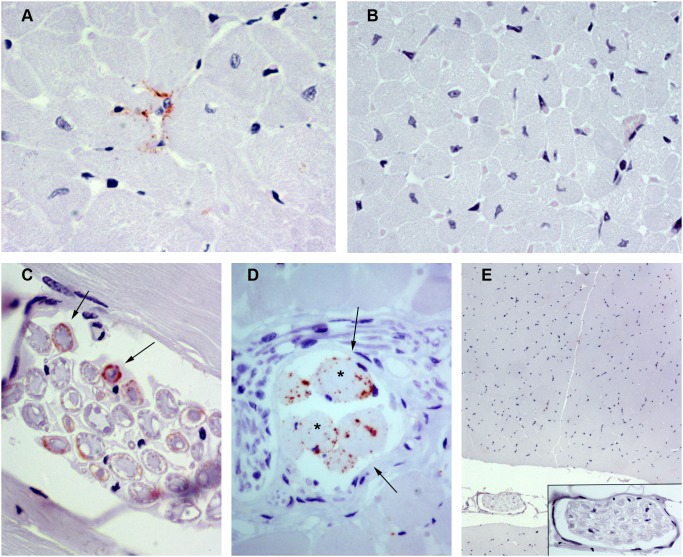
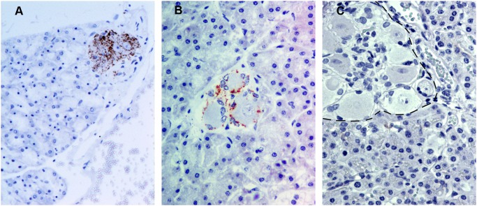
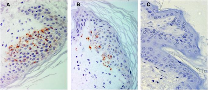
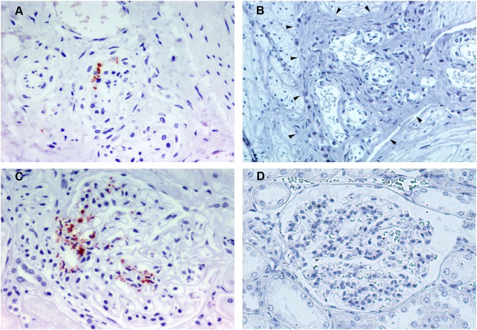
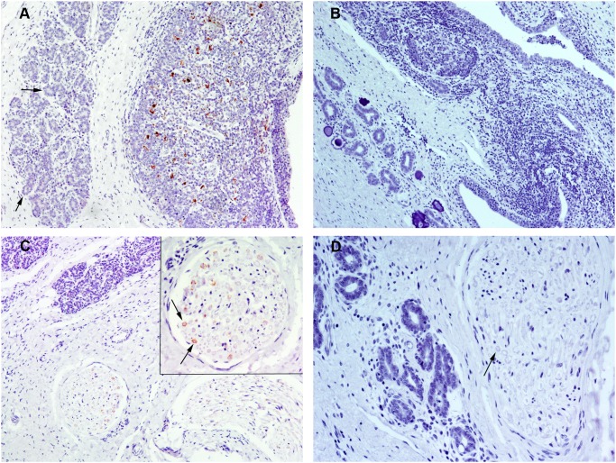
Accumulation of prion protein (PrPSc) in the central nervous system is the hallmark of transmissible spongiform encephalopathies. However, in some of these diseases such as scrapie or chronic wasting disease, the PrPSc can also accumulate in other tissues, particularly in the lymphoreticular system. In recent years, PrPSc in organs other than nervous and lymphoid have been described, suggesting that distribution of this protein in affected individuals may be much larger than previously thought. In the present study, 11 non-nervous/non-lymphoid organs from 16 naturally scrapie infected sheep in advanced stages of the disease were examined for the presence of PrPSc. Fourteen infected sheep were of the ARQ/ARQ PRNP genotype and 2 of the VRQ/VRQ, where the letters A, R, Q, and V represent the codes for amino-acids alanine, arginine, glutamine and valine, respectively. Adrenal gland, pancreas, heart, skin, urinary bladder and mammary gland were positive for PrPSc by immunohistochemistry and IDEXX HerdChek scrapie/BSE Antigen EIA Test in at least one animal. Lung, liver, kidney and skeletal muscle exhibited PrPSc deposits by immunohistochemistry only. To our knowledge, this is the first report regarding the presence of PrPSc in the heart, pancreas and urinary bladder in naturally acquired scrapie infections. In some other organs examined, in which PrPSc had been previously detected, PrPSc immunolabeling was observed to be associated with new structures within those organs. The results of the present study illustrate a wide dissemination of PrPSc in both ARQ/ARQ and VRQ/VRQ infected sheep, even when the involvement of the lymphoreticular system is scarce or absent, thus highlighting the role of the peripheral nervous system in the spread of PrPSc.


Conflict of interest statement

Competing Interests: The authors have declared that no competing interests exist.

Figures

Figure 1
Figure 1. Immunohistochemical detection of PrPSc in adrenal glands using L42 PrP antibody.
A) Adrenal medulla from a naturally scrapie-infected sheep (x40). Granular intracytoplasmic PrPSc immunolabeling is associated with chromaffin cells (arrow). B) Adrenal medulla from a naturally scrapie-infected sheep with an intense immunoreactivity (x20). Linear PrPSc immunolabeling can be observed also associated with chromaffin cells (arrows). C) Adrenal medulla from an uninfected control sheep in which no PrPSc immunolabeling is present (x10). D) Adrenal capsule and cortex from a naturally scrapie-infected sheep (x20). Perineuronal PrPSc immunolabeling is associated with neurons located beneath the capsule. E) Suprarenal ganglion from a naturally scrapie-infected sheep with abundant granular immunoreactivity for PrPSc (x5). A detailed image of the ganglion cells is shown in the insert picture in which PrPSc immunolabeling is observed mainly around ganglion cells.
Figure 2
Figure 2. Immunohistochemical detection of PrPSc in heart and skeletal muscle using L42 PrP antibody.
A) Heart from a naturally scrapie-infected sheep. PrPSc immunolabeling is located among myocardial myocytes (x63). B) Heart from an uninfected control sheep in which no PrPSc immunolabeling is present (x40). C) Cross section of a nerve fiber bundle located within a skeletal muscle sample from a naturally scrapie-infected sheep (x63). A periaxonal PrPSc immunolabeling can be observed in some nerve fibers (arrows). D) Skeletal muscle from a naturally scrapie-infected sheep in which PrPSc immunolabeling is associated with a neuromuscular spindle (cross section; x63). The neuromuscular spindle was identified by its typical structure: small groups of specialized striated muscle fibers (asterisks), which are thinner than regular muscle fibers, surrounded by a capsule of connective tissue (arows). E) Skeletal muscle from an uninfected control sheep in which no PrPSc immunolabeling is present (x10). A detailed image of a nerve fiber bundle immersed in the muscle is shown in the insert picture.
Figure 3
Figure 3. Immunohistochemical detection of PrPSc in pancreas using L42 PrP antibody.
A–B) Pancreas from naturally scrapie-infected sheep. PrPSc immunolabeling was detected only in relation to intrapancreatic ganglia neurons, showing a granular intracytoplasmic (A; x20) or a perineuronal (B; x40) labelling. C) Pancreas from an uninfected control sheep in which an intrapancreatic ganglion is indicated by the dotted line (x40). No PrPSc immunolabeling is present.
Figure 4
Figure 4. Immunohistochemical detection of PrPSc in skin.
A–B) Skin from a naturally scrapie-infected sheep with PrPSc immunolabeling located mainly at the epidermis. PrPSc was detected using L42 (A; x40) and F89 (B; x40) PrP monoclonal antibodies. C) Skin from an uninfected control sheep in which no PrPSc immunolabeling is present (x40).
Figure 5
Figure 5. Immunohistochemical detection of PrPSc in urinary bladder and kidney using L42 PrP antibody.
A) Urinary bladder muscularis from a naturally scrapie-infected sheep (x40). There is a weak PrPSc immunolabeling located in the connective tissue adjacent to the smooth muscle bundles, probably associated with vasculonervous elements. B) Urinary bladder muscularis from an uninfected control sheep in which no PrPSc immunolabeling is present (x20). The connective tissue (indicated by arrows head) with vasculonervous elements located between the smooth muscle can be clearly seen with low magnification. C) Kidney from a naturally scrapie-infected sheep. PrPSc immunolabeling is observed mainly at the vascular pole of a renal corpuscle (x40). D) A renal corpuscule of a kidney from an uninfected control sheep in which no PrPSc immunolabeling is present (x20).
Figure 6
Figure 6. Immunohistochemical detection of PrPSc in mammary gland using L42 PrP antibody.
A) Mammary gland from a sheep simultaneously infected by scrapie and by Maedi-visna infection causing a mastitis (x10). PrPSc immunolabeling is located within the lymphofollicular inflammatory lesions. In the mammary gland acini (arrows) next to the inflammatory lesions no PrPSc immunolabeling can be observed. B) Mammary gland from an uninfected control sheep with mastitis associated with Maedi-visna infection in which no PrPSc immunolabeling is present (x10). C) Mammary gland from a naturally scrapie-infected sheep (x10). PrPSc immunolabeling can be observed in the nerves immersed in the interlobular connective tissue (x10). A detailed image of a nerve fibers bundle in which a periaxonal PrPSc immunolabeling can be observed (arrows) is shown in the insert picture. D) Mammary gland from an uninfected control sheep in which no PrPSc immunolabeling is present (x20). Cross section of a nerve fiber is indicated by an arrow.
Figure 7
Figure 7. Immunohistochemical detection of PrPSc in lung and liver using L42 PrP antibody.
A) Lung from a sheep infected by scrapie with a concomitant verminous pneumonia (x10). PrPSc immunolabeling is located within the inflammatory lesions related to the lungworm infection (x10). Parasites can be observed in the alveoli (asterisk). B) Lung from an uninfected control sheep with verminous pneumonia in which no PrPSc immunolabeling is present (x10). Parasites can be observed in the alveoli (asterik). C) Liver from a sheep infecetd by scrapie with a concomitant trematode infection (x10). PrPSc immunolabeling is associated with a lymphofollicular inflammatory site around a bile duct. In the bile duct lumen adult forms of parasites can be observed (asterisk). D) Liver from an uninfected scrapie sheep with trematode infection in which no PrPSc immunolabeling is present (x10). In the bile duct lumen, adult forms of parasites can be observed (asterisk).

References

    1. Beekes M, McBride PA (2007) The spread of prions through the body in naturally acquired transmissible spongiform encephalopathies. FEBS J 274: 588–605. - PubMed
    1. Jeffrey M, Gonzalez L (2004) Pathology and pathogenesis of bovine spongiform encephalopathy and scrapie. Curr Top Microbiol Immunol 284: 65–97. - PubMed
    1. van Keulen LJ, Schreuder BE, Vromans ME, Langeveld JP, Smits MA (1999) Scrapie-associated prion protein in the gastrointestinal tract of sheep with natural scrapie. J Comp Pathol 121: 55–63. - PubMed
    1. Andreoletti O, Berthon P, Marc D, Sarradin P, Grosclaude J, et al. (2000) Early accumulation of PrP(Sc) in gut-associated lymphoid and nervous tissues of susceptible sheep from a Romanov flock with natural scrapie. J Gen Virol 81: 3115–3126. - PubMed
    1. Tuo W, ÓRourke K, Zhuang D, Cheevers W, Spraker T (2002) Pregnancy status and fetal prion genetics determine PrPSc accumulation in placentomes of scrapie infected sheep. PNAS 99: 6310–6315. - PMC - PubMed

Publication types

MeSH terms