. 2014 Jun 27;289(26):18514-25.
doi: 10.1074/jbc.M114.559930. Epub 2014 May 14.

SCF-FBXO31 E3 ligase targets DNA replication factor Cdt1 for proteolysis in the G2 phase of cell cycle to prevent re-replication

Affiliations

SCF-FBXO31 E3 ligase targets DNA replication factor Cdt1 for proteolysis in the G2 phase of cell cycle to prevent re-replication

Pegah Johansson et al. J Biol Chem. .

Abstract

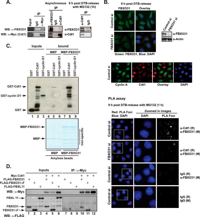
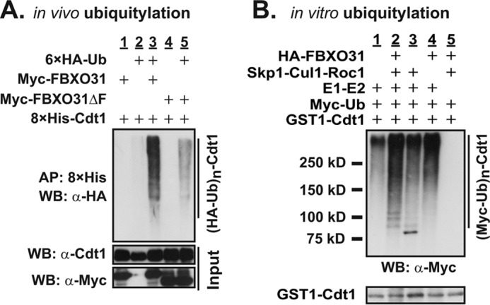
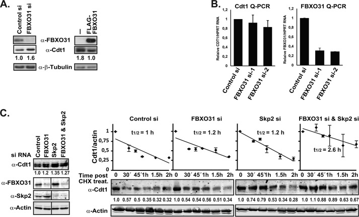
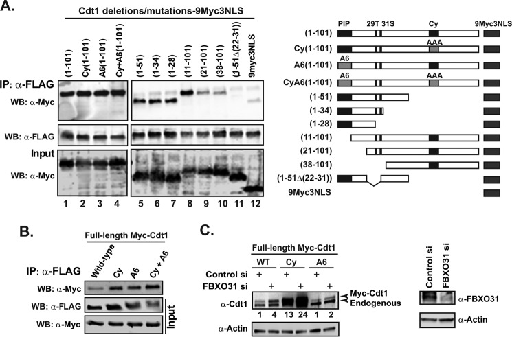
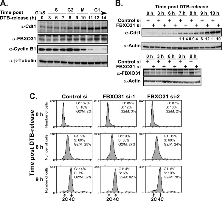
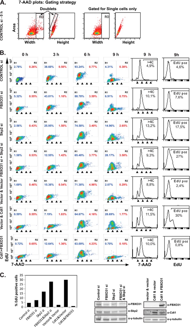
FBXO31 was originally identified as a putative tumor suppressor gene in breast, ovarian, hepatocellular, and prostate cancers. By screening a set of cell cycle-regulated proteins as potential FBXO31 interaction partners, we have now identified Cdt1 as a novel substrate. Cdt1 DNA replication licensing factor is part of the pre-replication complex and essential for the maintenance of genomic integrity. We show that FBXO31 specifically interacts with Cdt1 and regulates its abundance by ubiquitylation leading to subsequent degradation. We also show that Cdt1 regulation by FBXO31 is limited to the G2 phase of the cell cycle and is independent of the pathways previously described for Cdt1 proteolysis in S and G2 phase. FBXO31 targeting of Cdt1 is mediated through the N terminus of Cdt1, a region previously shown to be responsible for its cell cycle regulation. Finally, we show that Cdt1 stabilization due to FBXO31 depletion results in re-replication. Our data present an additional pathway that contributes to the FBXO31 function as a tumor suppressor.

Keywords: Cdt1; Cell Cycle; DNA Replication; E3 Ubiquitin Ligase; FBXO31; Genomic Instability; Protein Degradation.


Figures

FIGURE 1.
FIGURE 1.
FBXO31 interacts with Cdt1. A, HEK293T cells were transfected with Myc-Cdt1 expression construct. Cell lysates were immunoprecipitated (IP) with either mouse IgG control or anti-Myc antibody. Input and IP samples were Western-blotted (WB) with anti-Myc or anti-FBXO31 antibodies (left panel). For endogenous immunoprecipitation, either asynchronous HeLa cells or cells synchronized with a double-thymidine block and harvested 9 h after release were used. The synchronized cells were treated with MG132 for 1 h before harvest. Total cell extract was subjected to immunoprecipitation using anti-Cdt1, anti-FBXO31 antibodies, or rabbit IgG. The immunoprecipitates were analyzed by Western blotting with anti-FBXO31 and anti-Cdt1 antibodies (right panel). B, HeLa cells were transiently transfected with control or FBXO31 siRNA (siRNA-1), synchronized with a double-thymidine block, and fixed onto coverslips 9 h after release. Samples were treated with MG132 for 1 h before fixation where indicated on the figure. Cells were probed with anti-FBXO31, anti-Cdt1, and anti-cyclin A antibodies or mouse (M) or rabbit (R) IgG alone or in combination as indicated on the figure. Antibodies were either detected by fluorescently labeled secondary antibodies or a proximity ligation assay kit as marked. C, GST, GST-Cdt1, or GST-cyclin D1 proteins expressed and purified from bacteria were incubated with bacterially-expressed MBP or MBP-FBXO31 fusion proteins attached to amylose resin. The complexes were washed and eluted in 1× protein-loading buffer and either analyzed by Western blotting with anti-GST antibody (upper image) or resolved by SDS-PAGE and visualized by colloidal Coomassie staining (lower image). Note a low level of nonspecific GST-Cdt1 binding to MBP-beads in lane 5. D, HEK293T cell lysates expressing FLAG-FBXO31, FLAG-FBXO31ΔF, or FLAG-FBXL11 either alone or together with Myc-Cdt1 were immunoprecipitated with anti-Myc antibody. Inputs and IP samples were Western-blotted with anti-Myc or anti-FLAG antibodies. Note different exposures of the same blot with input and IP samples probed with anti-FLAG antibody were used for clarity.
FIGURE 2.
FIGURE 2.
SCF-FBXO31 ubiquitylates Cdt1. A, in vivo ubiquitylation assay was performed on HEK293T cells transfected with constructs expressing tagged proteins as shown and incubated with 10 μm MG132 for 4 h before harvesting. Whole cell extracts (bottom panel) and 8×His-Cdt1 affinity-purified (AP) on nickel-nitrilotriacetic acid Spin Columns (Qiagen) under denaturing conditions were Western-blotted (WB) with antibodies as shown. Polyubiquitylated Cdt1 appears as a ladder in lane 3 (top blot). B, for in vitro ubiquitylation assay, SCF complexes were affinity-purified with anti-HA-conjugated agarose beads from HEK293T cells expressing HA-tagged Skp1, Cul1, and Roc1 with or without FBXO31 and incubated with GST-Cdt1, E1 (UBE1), E2 (UbcH5a and UbcH8), ubiquitylation buffer, Myc-ubiquitin, and Cdk2/cyclin A. The reaction was stopped by the addition of 1× protein-loading buffer, and samples were Western-blotted with antibodies as shown. Polyubiquitylated-Cdt1 appears as a ladder in lane 2 (top blot).
FIGURE 3.
FIGURE 3.
FBXO31 regulates Cdt1 protein stability. A, H1299 cells were transfected with FBXO31 (siRNA-1) or control siRNA and Western-blotted with anti-FBXO31, anti-Cdt1, and anti-β-tubulin (loading control) antibodies. Additionally, HEK293T and HEK293T cells overexpressing FLAG-FBXO31 were Western-blotted with anti-FLAG, anti-Cdt1, and anti-β-tubulin (loading control) antibodies. Cdt1 protein levels were then quantified using the ImageJ software and normalized against β-tubulin. B, asynchronous HeLa cells were transiently transfected with control or FBXO31 siRNAs, and Cdt1 and FBXO31 mRNA levels were measured using RT-qPCR assay and normalized against HPRT1 mRNA. C, asynchronous HeLa cells were transiently transfected with control, FBXO31 (siRNA-2), Skp2 or FBXO31 (siRNA-2), and Skp2 siRNA, subsequently treated with cyclohexamide (CHX; 100 μm) and harvested at the indicated time points for Western blot analysis using anti-Cdt1 and anti-actin antibodies. Protein extracts from the zero time point were also analyzed on Western blots with anti-FBXO31 and anti-Skp2 antibodies. Fluorescently labeled secondary antibodies were used, and the blots were scanned on a Li-Cor Odyssey scanner. Cdt1 protein levels were then quantified using the ImageJ software and normalized against actin. The 0 time point for each siRNA treatment was set to 1. Half-life analysis is based on two independent experiments. Note that a lane is taken out of the FBXO31, Skp2, and actin blots on the left where marked on the image, but they each represent the same blot and exposure.
FIGURE 4.
FIGURE 4.
FBXO31 interacts with Cdt1 N terminus and stabilizes Cdt1 Cy and A6 mutants. A, HEK293T cells stably expressing FLAG-FBXO31 were transfected with Cdt1 N-terminal constructs fused to 9myc3NLS. Cell lysates were IP with an anti-FLAG antibody. Input and IP samples were Western-blotted (WB) with anti-Myc or anti-FLAG antibodies. A schematic of the Cdt1 N-terminal constructs is illustrated below the figure. The 29T amino acid in the Cdt1 protein is a phosphorylation target closely located to Ser-31, which is also a potential phosphorylation target (47). B, HEK293T cells stably expressing FLAG-FBXO31 were transfected with constructs expressing full-length Myc tagged Cdt1 proteins (wild type, Cy, A6, or CyA6). Cell lysates were immunoprecipitated with an anti-FLAG antibody. Input and IP samples were Western-blotted with anti-Myc or anti-FLAG antibodies. C, HeLa cells were first transfected with control or FBXO31 siRNA (siRNA-2) on day 1, and then a control or FBXO31 siRNA set was transfected with either wild type, Cy, or A6-Cdt1 expression constructs on day 2. The cells were harvested 48 h post-transfection and analyzed by Western blotting with anti-Cdt1, anti-FBXO31, and anti-actin antibodies. The Myc-Cdt1 protein levels were quantified using the ImageJ software and normalized against actin.
FIGURE 5.
FIGURE 5.
FBXO31 destabilizes Cdt1 in G2. A, HeLa cells were synchronized by double-thymidine block, harvested at the indicated time points after release from the block, and analyzed by Western blotting with anti-Cdt1, anti-FBXO31, anti-cyclin B1, and anti-β-tubulin antibodies. B, HeLa cells were transfected with control or FBXO31 siRNAs (siRNA-1 shown for panel C), synchronized with a double-thymidine block (DTB), and harvested at the indicated time points after release. The cells were analyzed by Western blotting with anti-Cdt1, anti-FBXO31, and anti-actin antibodies. Cdt1 protein levels were quantified using the ImageJ software and normalized against actin, and the cells were stained with a cell cycle marker (7-AAD) and analyzed by flow cytometry (C).
FIGURE 6.
FIGURE 6.
FBXO31 depletion induces re-replication. HeLa cells were transiently transfected with control, FBXO31, Skp2 or FBXO31 (siRNA-2), and Skp2 siRNAs 72 h before harvest. Cells were also transfected with empty vectors, Cdt1 and vector, or Cdt1 and FBXO31 expression plasmids. The transfected cells were synchronized by a double-thymidine block and harvested at the indicated time points after release from the block. All the samples were pulsed with EdU for 30 min before harvest. The samples were analyzed by flow cytometry. A, dot plots of the 7-AAD area versus 7-AAD width were used to gate for single cells only as shown for HeLa cells transfected with control si at time 0. B, the EdU profile and cell cycle analysis data are presented as dot plots. The arrowheads indicate cells with 2C, 4C, 6C, and 8C DNA content. The EdU and cell cycle profile (7-AAD) of cells at 9 h are also presented as histograms in the right panels. C, bar graph showing the relative percentage of EdU positive cells. Western blotting was carried out with anti-FBXO31, anti-Skp2, anti-Cdt1 and anti-γ-tubulin antibodies.

Similar articles

Cited by

References

    1. Härkönen P., Kyllönen A. P., Nordling S., Vihko P. (2005) Loss of heterozygosity in chromosomal region 16q24.3 associated with progression of prostate cancer. Prostate 62, 267–274 - PubMed
    1. Kumar R., Neilsen P. M., Crawford J., McKirdy R., Lee J., Powell J. A., Saif Z., Martin J. M., Lombaerts M., Cornelisse C. J., Cleton-Jansen A. M., Callen D. F. (2005) FBXO31 is the chromosome 16q24.3 senescence gene, a candidate breast tumor suppressor, and a component of an SCF complex. Cancer Res. 65, 11304–11313 - PubMed
    1. Launonen V., Mannermaa A., Stenbäck F., Kosma V. M., Puistola U., Huusko P., Anttila M., Bloigu R., Saarikoski S., Kauppila A., Winqvist R. (2000) Loss of heterozygosity at chromosomes 3, 6, 8, 11, 16, and 17 in ovarian cancer: correlation to clinicopathological variables. Cancer Genet. Cytogenet. 122, 49–54 - PubMed
    1. Lin Y. W., Lee I. N., Chen C. H., Huang G. T., Lee H. S., Lee P. H., Lu F. J., Sheu J. C. (2001) Deletion mapping of chromosome 16q24 in hepatocellular carcinoma in Taiwan and mutational analysis of the 17-β-HSD gene localized to the region. Int. J. Cancer 93, 74–79 - PubMed
    1. Miller B. J., Wang D., Krahe R., Wright F. A. (2003) Pooled analysis of loss of heterozygosity in breast cancer: a genome scan provides comparative evidence for multiple tumor suppressors and identifies novel candidate regions. Am. J. Hum. Genet. 73, 748–767 - PMC - PubMed

Publication types