Review
. 2014 Jun;11(5):811-22.
doi: 10.1513/AnnalsATS.201312-446FR.

Management of acute right ventricular failure in the intensive care unit

Affiliations
Review

Management of acute right ventricular failure in the intensive care unit

Corey E Ventetuolo et al. Ann Am Thorac Soc. 2014 Jun.

Abstract

Right ventricular (RV) failure occurs when the RV fails to maintain enough blood flow through the pulmonary circulation to achieve adequate left ventricular filling. This can occur suddenly in a previously healthy heart due to massive pulmonary embolism or right-sided myocardial infarction, but many cases encountered in the intensive care unit involve worsening of compensated RV failure in the setting of chronic heart and lung disease. Management of RV failure is directed at optimizing right-sided filling pressures and reducing afterload. Due to a lower level of vascular tone, vasoactive medications have less salient effects on reducing vascular resistance in the pulmonary than in the systemic circulation. Successful management requires reversal of any conditions that heighten pulmonary vascular tone and the use of selective pulmonary vasodilators at doses that do not induce systemic hypotension or worsening of oxygenation. Systemic systolic arterial pressure should be kept close to RV systolic pressure to maintain RV perfusion. When these efforts fail, the judicious use of inotropic agents may help improve RV contractility enough to maintain cardiac output. Extracorporeal life support is increasingly being used to support patients with acute RV failure who fail to respond to medical management while the underlying cause of their RV failure is addressed.

Keywords: critical care; pulmonary hypertension; right-sided heart failure.


Figures

Figure 1.
Figure 1.
Drop in mean vascular pressure between different segments of the arterial and venous portions of the isolated dog lung as measured by micropuncture technique. Note the small decrease in pressure as pulmonary arterial diameter decreases compared with the large drop in resistance between precapillary arterioles and post-capillary venuoles. Adapted by permission from Reference .
Figure 2.
Figure 2.
Differences between right ventricular (RV) and left ventricular (LV) response to increasing afterload (left panel) and increasing preload (right panel). RV stroke volume falls sharply as mean vascular pressure () is increased from 20 to 30 mm Hg in the pulmonary artery, but LV stroke volume stays fairly constant as mean aortic pressure is increased from 100 to 140 mm Hg. In contrast, LV stroke work increases rapidly as left atrial pressure is raised from 10 to 20 cm H2O, while the increase in RV stroke work in response to the same elevation or right atrial pressure is much more modest. Reproduced by permission from Reference .
Figure 3.
Figure 3.
The position of the interventricular septum during the cardiac cycle is determined by the difference between right ventricular (RV) and left ventricular (LV) pressure. Under normal conditions, LV end-diastolic pressure is greater than RV end-diastolic pressure, and the septum bows toward the RV during diastole. As the RV fails, RV end-diastolic pressure begins to exceed that of the left ventricle (LV) and the septum bows toward the LV during diastole forming a “D”-shaped pattern and impaired LV filling (left panel). When RV failure occurs due to elevated pulmonary vascular resistance, the combination of high RV systolic pressure and decreased LV filling may lead to near obliteration of the LV at end systole (right panel).
Figure 4.
Figure 4.
Effect of oxygen tension and acidemia on pulmonary vasoconstriction. (A) Pulmonary vasoconstriction increases as alveolar O2 tension (PAO2) falls while keeping mixed venous oxygenation constant (mixed venous Po2 indicated for each solid line from 60 to 10 mm Hg). The combined effect of allowing PAO2 and mixed venous Po2 to fall is shown by the dashed line where PAO2 and mixed venous oxygen saturation O2 are the same (34). Maximal pulmonary vasoconstrictor response was defined as the difference between baseline pulmonary artery pressure when ventilating with an FiO2 of 0.21 and perfusate FIO2 of 0.06, and the pulmonary artery pressure when both the inspired and the perfusate FiO2 was zero. The pressure response at all other combinations of inspired and perfusate FiO2 were expressed as a percent of this 10 maximum (%Rmax). (B) Pulmonary vasoconstriction in newborn calves as a function of inspired Po2 under conditions of different levels of arterial blood pH. Hypoxic pulmonary vasoconstriction is increased and occurs at a higher level of inspired O2 as arterial pH is decreased. PVR = pulmonary vascular resistance. Reproduced by permission from Reference .
Figure 5.
Figure 5.
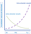
Pulmonary vascular resistance in intra- and extra-alveolar vessels relative to lung volume. Interstitial pressure becomes more negative during lung expansion in the spontaneously breathing patient, favoring enlargement of most pulmonary vessels and a fall in their resistance to blood flow. Intra-avleolar vessels, however, become compressed by expansion of surrounding alveoli during lung inflation, leading to increased vascular resistance in these vessels. Total pulmonary vascular resistance is lowest at functional residual capacity, where vascular resistance in both intra- and extra-alveolar vessels is intermediate. TLC = total lung capacity. Reproduced by permission from Reference .
Figure 6.
Figure 6.
Cycle of right ventricular (RV) decompensation. RV dysfunction begins with excessive increases in preload or afterload, or injury that results in decreased contractility. Tachycardia and increased stroke work lead to increased RV free wall tension, resulting in increased oxygen demand. Eventually, cardiac output begins to fall, leading to systemic hypotension. RV ischemia ensues from the combination of increased RV work load, free wall tension, and decreased coronary perfusion. Inadequate delivery of oxygen to the RV myocardium worsens myocyte contractility, leading to greater RV dilation and wall tension (left-sided arrow) and decreased O2 delivery, due to systemic hypotension, lower mixed venous oxygen saturation, and increased right-to-left-side intracardiac shunting as RV pressure rises (right-sided arrow). The pulmonary vascular disease associated with pulmonary arterial hypertension (PAH) poses a particularly difficult situation for RV function, because it increases RV afterload while decreasing oxygenation of pulmonary venous blood leading to systemic (including coronary arterial) hypoxemia. PFO = patent foramen ovale. Reproduced by permission from Reference .
Figure 7.
Figure 7.
Ratio of blood flow during systole to blood flow during diastole in the right coronary artery (RCA) as a function of right ventricular (RV) systolic pressure in patients with pulmonary hypertension. When RV systolic pressure is near normal levels, the amount of RCA blood flow is similar during systole and diastole. As RV systolic pressure increases, RCA blood flow during systole falls. Blood flow in the RCA during systole is inversely proportional to RV systolic pressure, such that RCA blood flow during systole approaches zero as RV systolic pressure approaches systemic levels. Reproduced by permission from Reference .
Figure 8.
Figure 8.
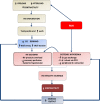
Approach to management of acute right ventricular (RV) failure. Patients should be assessed for acute cause of increased RV afterload or decreased contractility, such as pulmonary embolism or right-sided infarction. If no readily reversible cause is identified, efforts should be directed at optimizing RV preload and reducing RV afterload. The latter should include reversal of factors known to increase pulmonary vascular resistance and then the use of selective pulmonary vasodilator drugs. Metabolic conditions that reduce cardiac contractility, such as sepsis and acidemia, should be addressed. Systemic arterial pressure should be kept above RV systolic pressure to maintain RV perfusion. If these efforts fail, ionotropic agents can be tried to improve RV contractility. Measures to correct metabolic abnormalities and reduce RV dilation will also aid RV contractility. Extracorporeal life support should be considered when medical therapy is unsuccessful in a patient who has a reversible cause of RV failure or who is being prepared for lung transplantation. ECLS = extracorporeal life support; PE = pulmonary embolism; PVR = pulmonary vascular resistance.

References

    1. Bhattacharya J, Staub NC. Direct measurement of microvascular pressures in the isolated perfused dog lung. Science. 1980;210:327–328. - PubMed
    1. Dell’Italia LJ. The right ventricle: anatomy, physiology, and clinical importance. Curr Probl Cardiol. 1991;16:653–720. - PubMed
    1. Greyson CR. Pathophysiology of right ventricular failure. Crit Care Med. 2008;36(1) Suppl:S57–S65. - PubMed
    1. James TN. Anatomy of the crista supraventricularis: its importance for understanding right ventricular function, right ventricular infarction and related conditions. J Am Coll Cardiol. 1985;6:1083–1095. - PubMed
    1. Pouleur H, Lefèvre J, Van Mechelen H, Charlier AA. Free-wall shortening and relaxation during ejection in the canine right ventricle. Am J Physiol. 1980;239:H601–H613. - PubMed

Publication types

LinkOut - more resources