. 2014 May 27;111(21):7683-8.
doi: 10.1073/pnas.1310972111. Epub 2014 May 14.

Protein kinase D1 is essential for Ras-induced senescence and tumor suppression by regulating senescence-associated inflammation

Affiliations

Protein kinase D1 is essential for Ras-induced senescence and tumor suppression by regulating senescence-associated inflammation

Pan Wang et al. Proc Natl Acad Sci U S A. .

Abstract

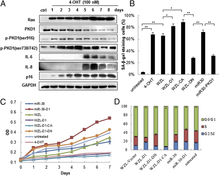
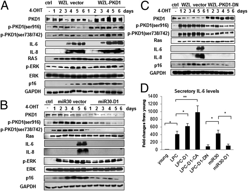
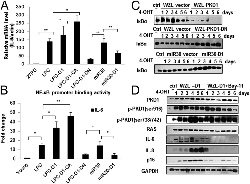
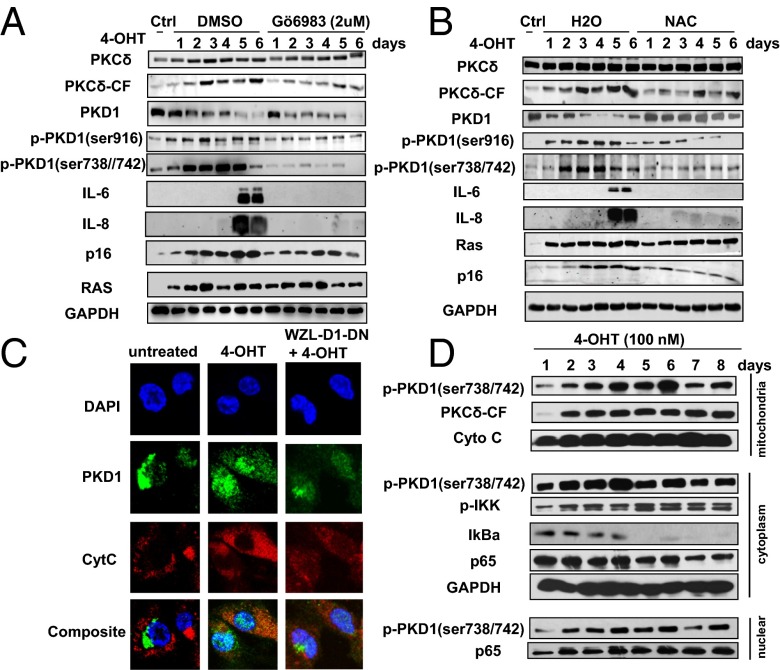
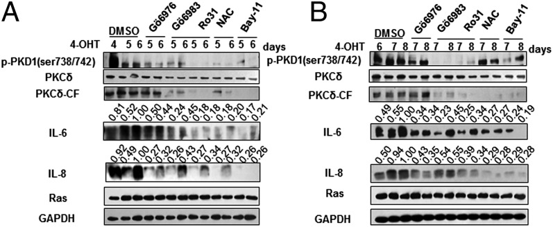
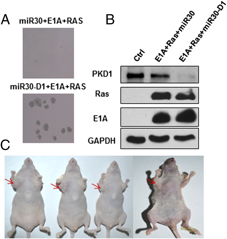
Oncogene-induced senescence (OIS) is an initial barrier to tumor development. Reactive oxygen species (ROS) is critical for oncogenic Ras OIS, but the downstream effectors to mediate ROS signaling are still relatively elusive. Senescent cells develop a senescence-associated secretory phenotype (SASP). However, the mechanisms underlying the regulation of the SASP are largely unknown. Here, we identify protein kinase D1 (PKD1) as a downstream effector of ROS signaling to mediate Ras OIS and SASP. PKD1 is activated by oncogenic Ras expression and PKD1 promotes Ras OIS by mediating inflammatory cytokines interleukin-6 (IL-6) and interleukin-8 (IL-8) via modulation of NF-κB activity. We demonstrate that ROS-protein kinase Cδ (PKCδ)-PKD1 axis is essential for the establishment and maintenance of IL-6/IL8 induction. In addition, ablation of PKD1 causes the bypass of Ras OIS, and promotes cell transformation and tumorigenesis. Together, these findings uncover a previously unidentified role of ROS-PKCδ-PKD1 pathway in Ras OIS and SASP regulation.


Conflict of interest statement

The authors declare no conflict of interest.

Figures

Fig. 1.
Fig. 1.
PKD1 is activated during Ras-induced senescence and is required for Ras OIS. (A) PKD1 is activated by Ras induction. ER:Ras IMR90 cells were given 100 nM 4-OHT for the indicated days; fresh medium with 4-OHT was changed every other day. Cell lysates were collected every day for total eight days, and then analyzed for expression of the indicated proteins. (BD) PKD1 overexpression enhances Ras OIS, whereas PKD1 silencing prevents Ras OIS. ER:Ras IMR90 cells expressing the indicated genes and shRNAs were induced to express Ras for 6 d. (B) Cells were stained for SA-β-gal. The percentage of cells positive for SA-β-gal in each sample was shown. At least 300 cells were counted for each sample. Error bars represent means + SD (n = 3) *P < 0.05, **P < 0.01. (C) Growth curves were determined by 3-(4,5-dimethylthiazol-2-yl)-2,5-diphenyltetrazolium bromide assay. (D) Cell cycle was measured. Values are mean ± SD of triplicate points from a representative experiment (n = 3), which was repeated three times with similar results.
Fig. 2.
Fig. 2.
PKD1 mediates Ras OIS via modulation of IL-6/IL-8 induction. (AC) PKD1 overexpression promotes IL-6/IL-8 expression, and PKD1 silencing abolishes IL-6/IL-8 induction. ER:Ras IMR90 cells stably transduced with WZL-PKD1 (A), miR30-PKD1 (B), and WZL-PKD1-DN (C) were given 4-OHT for the indicated days. Cell lysates were then subjected to Western blot analysis for the indicated proteins. Data are representative of three independent experiments. (D) Supernatants collected from above cells at day 6 after Ras induction were assessed secretory levels of IL-6 measured by ELISA. Three independent experiments were analyzed. Error bars represent means + SD (n = 3) *P < 0.05, **P < 0.01.
Fig. 3.
Fig. 3.
PKD1 regulates IL-6/IL-8 at mRNA level and depends on NF-κB activity in Ras OIS. (A) PKD1 regulates IL-6 at mRNA level. Total mRNA were extracted from ER:Ras IMR90 cells expressing indicated genes and shRNAs after 6 d Ras induction. Relative mRNA level of IL-6 was determined by quantitative PCR. mRNA levels in young IMR90 cells are considered as control. (B) PKD1 affects NF-κB binding activity to the promoter of IL-6 gene. ER:Ras IMR90 cells expressing indicated genes and shRNAs were given 4-OHT for 6 d, and then the lysates were analyzed by ChIP assay using an antibody against NF-κB. NF-κB binding to the IL-6 promoters in the indicated stable transfectants is represented relative to mouse IgG binding and is normalized to young IMR90 cells binding for each other. Error bars represent means + SD (n = 3) *P < 0.05, **P < 0.01 in A and B. (C) PKD1 affects IκBα degradation. ER:Ras IMR90 cells expressing WZL-PKD1, WZL-PKD1-DN, and miR30-PKD1, respectively, were given 4-OHT for the indicated times, and then subjected to Western blot to detect IκBα protein levels. (D) NF-κB inhibition abolishes PKD1-mediated IL-6/IL-8 expression. ER:Ras IMR90 cells expressing WZL-PKD1 were treated with solvent DMSO or IκB kinases inhibitor BAY 11-7082 (5 μM) in the presence of 4-OHT for the indicated days, then analyzed for expression of indicated proteins.
Fig. 4.
Fig. 4.
ROS-PKCδ is upstream signaling to activate PKD1 in Ras OIS. (A) PKCδ inhibition blocks PKD1 activation and IL-6/IL-8 induction. ER:Ras IMR90 cells were given 4-OHT in the presence of solvent DMSO or Gö6983 (2 μM) for the indicated times; fresh medium with 4-OHT and Gö6983 was changed every other day. Cell lysates were then subjected to Western blot analysis for the indicated proteins. (B) Removal of ROS inhibits PKCδ-PKD1 activation and blocks IL-6/IL-8 induction. ER:Ras IMR90 cells treated with solvent or NAC (10 mM) for indicated times were subjected to Western blot analysis. (C) PKD1 translocates to the mitochondria. ER:Ras IMR90 cells and PKD1-DN–infected cells were seeded on glass coverslips and cultured with or without 4-OHT for 4 d, and then stained with phospho-PKD1 (ser738/742), green; anti-cytochrome c, red; and nuclei, DAPI, blue. (D) The coincidence of the kinetics of PKD1 activation, IKK complex phosphorylation, IκBα degradation, and p65 translocation to nucleus after Ras induction. ER:Ras IMR90 cells were cultured with 4-OHT and collected in the mitochondrial isolation buffer at indicated time; then, mitochondrial, cytoplasmic, and nuclear fractions were isolated and subjected to Western blot for the indicated proteins.
Fig. 5.
Fig. 5.
ROS-PKCδ-PKD1 signaling is required for the initiation and maintenance of IL-6/IL-8 induction. (A) Early interruption of this axis suppresses the initial induction of IL-6/IL-8. ER:Ras IMR90 cells were induced to express Ras for 4 d, then indicated inhibitors were added for another 1 or 2 d in the presence of 4-OHT. The indicated proteins were analyzed by Western blot. (B) Late intervention of this pathway inhibits the sustained production of IL-6/IL-8. ER:Ras IMR90 cells were cultured with 4-OHT for 6 d, then indicated inhibitors were added for another 1 or 2 d in the presence of 4-OHT. The indicated proteins were subjected to Western blot analysis. BAY 11-7082–treated cells serve as positive control. The densitometry data were analyzed by ImageJ software and normalized to the highest signal in the corresponding row.
Fig. 6.
Fig. 6.
PKD1 deficiency promotes cell transformation and tumorigenesis. (A) Normal IMR90 cells were transduced with Ras, E1A, and miR30 vector or miR30-D1. The two stable cell lines grew in soft agar as described. (B) Levels of PKD1, Ras, and E1A in the above two cell lines were determined by Western blot. (C) The transformation cells (miR30-D1+Ras+E1A) can form tumors in nude mice. About 5 × 10⁶ cells were collected, and resuspended in 0.1 mL of PBS, and then injected into mice. Control and transformed cells were injected to the left and right forelimb armpit, respectively. Tumor development was photographed at 5 wk after injection.

References

    1. Kuilman T, Michaloglou C, Mooi WJ, Peeper DS. The essence of senescence. Genes Dev. 2010;24(22):2463–2479. - PMC - PubMed
    1. Collado M, Serrano M. Senescence in tumours: Evidence from mice and humans. Nat Rev Cancer. 2010;10(1):51–57. - PMC - PubMed
    1. Serrano M, Lin AW, McCurrach ME, Beach D, Lowe SW. Oncogenic ras provokes premature cell senescence associated with accumulation of p53 and p16INK4a. Cell. 1997;88(5):593–602. - PubMed
    1. Lee AC, et al. Ras proteins induce senescence by altering the intracellular levels of reactive oxygen species. J Biol Chem. 1999;274(12):7936–7940. - PubMed
    1. Wu C, Miloslavskaya I, Demontis S, Maestro R, Galaktionov K. Regulation of cellular response to oncogenic and oxidative stress by Seladin-1. Nature. 2004;432(7017):640–645. - PubMed

Publication types