. 2014 Jul;155(7):2391-401.
doi: 10.1210/en.2013-1985. Epub 2014 May 14.

Defective transport of the obesity mutant PC1/3 N222D contributes to loss of function

Affiliations

Defective transport of the obesity mutant PC1/3 N222D contributes to loss of function

Yogikala Prabhu et al. Endocrinology. 2014 Jul.

Abstract

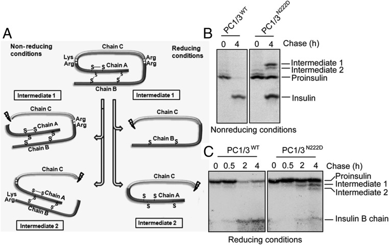
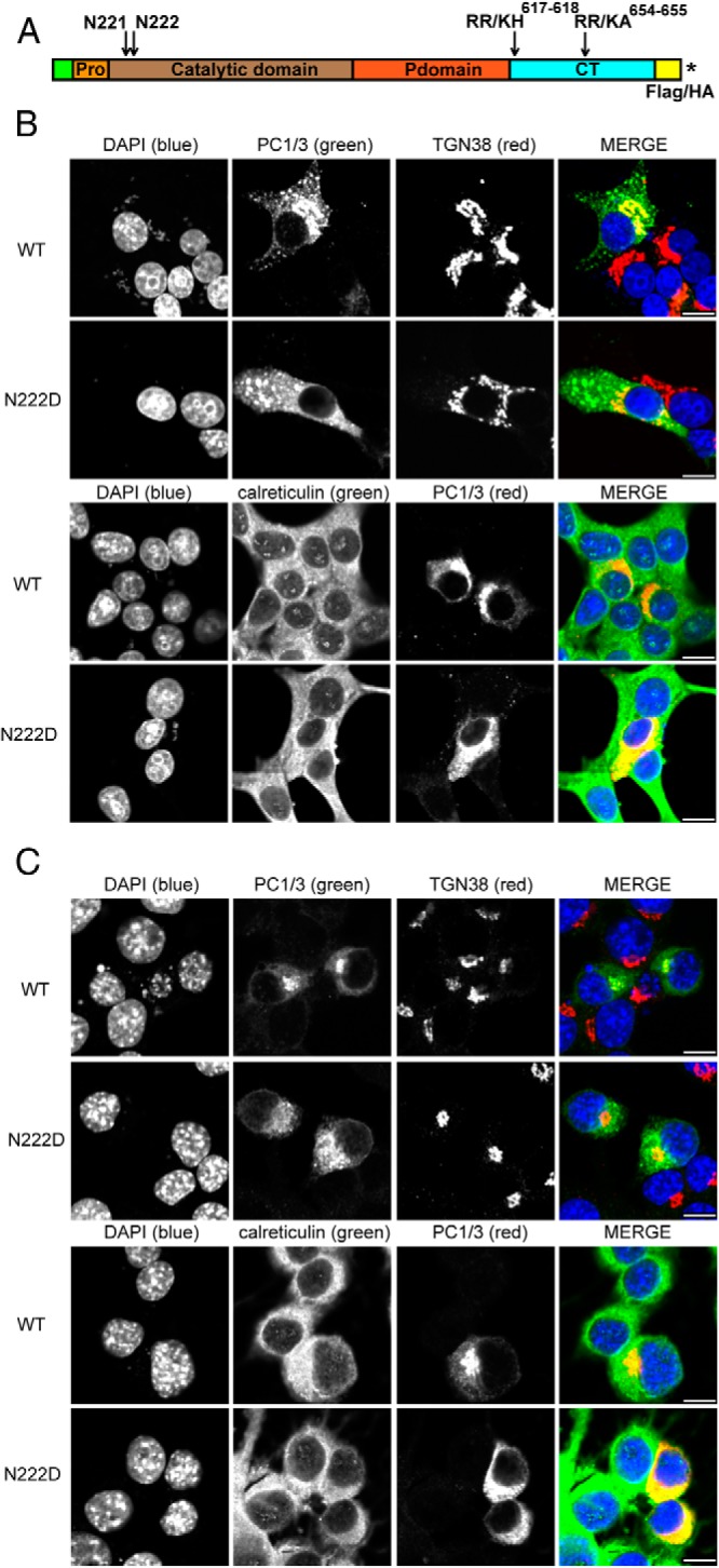
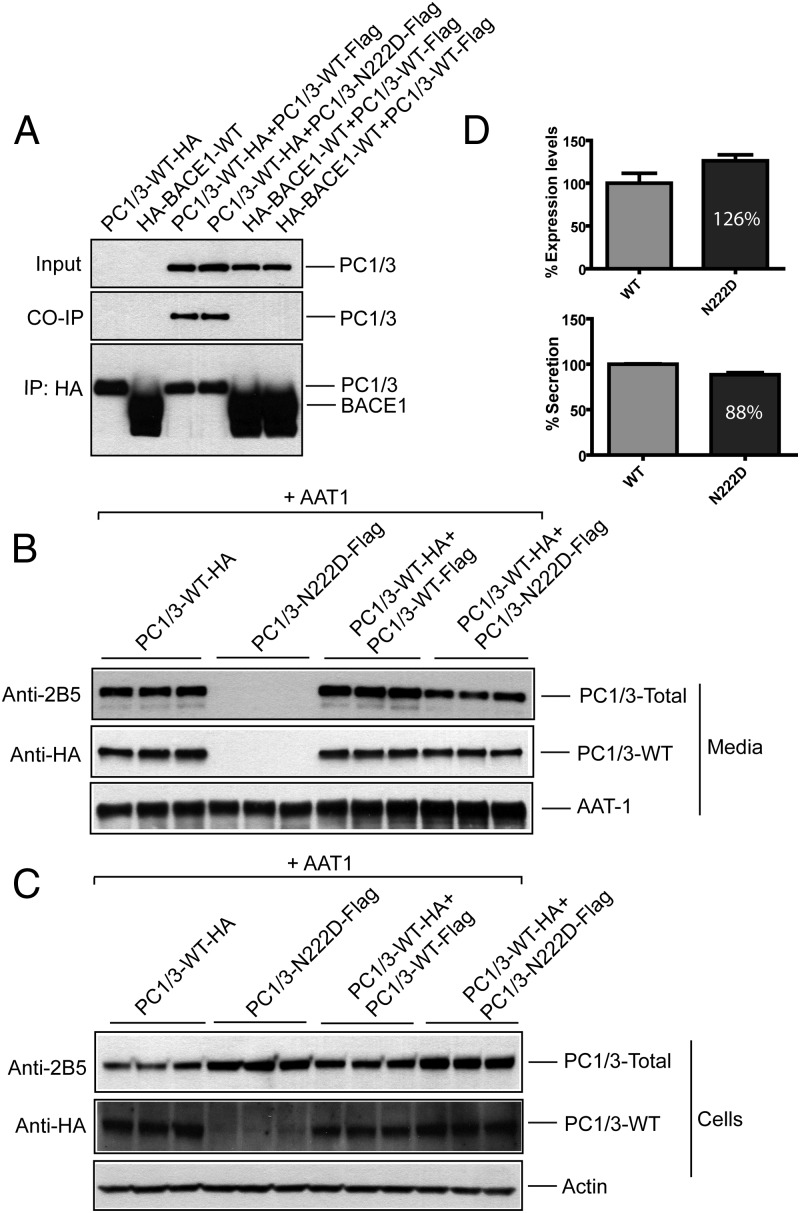
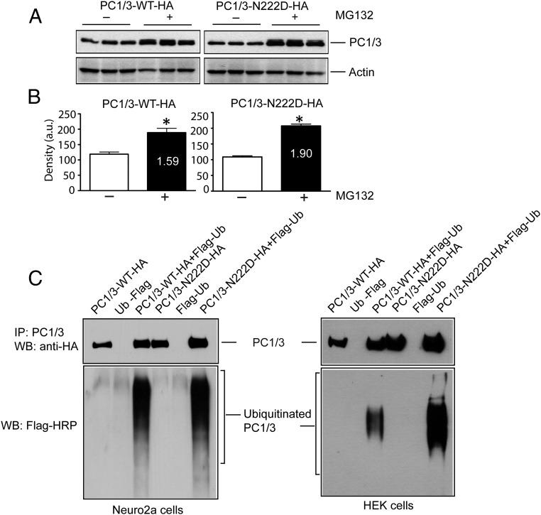
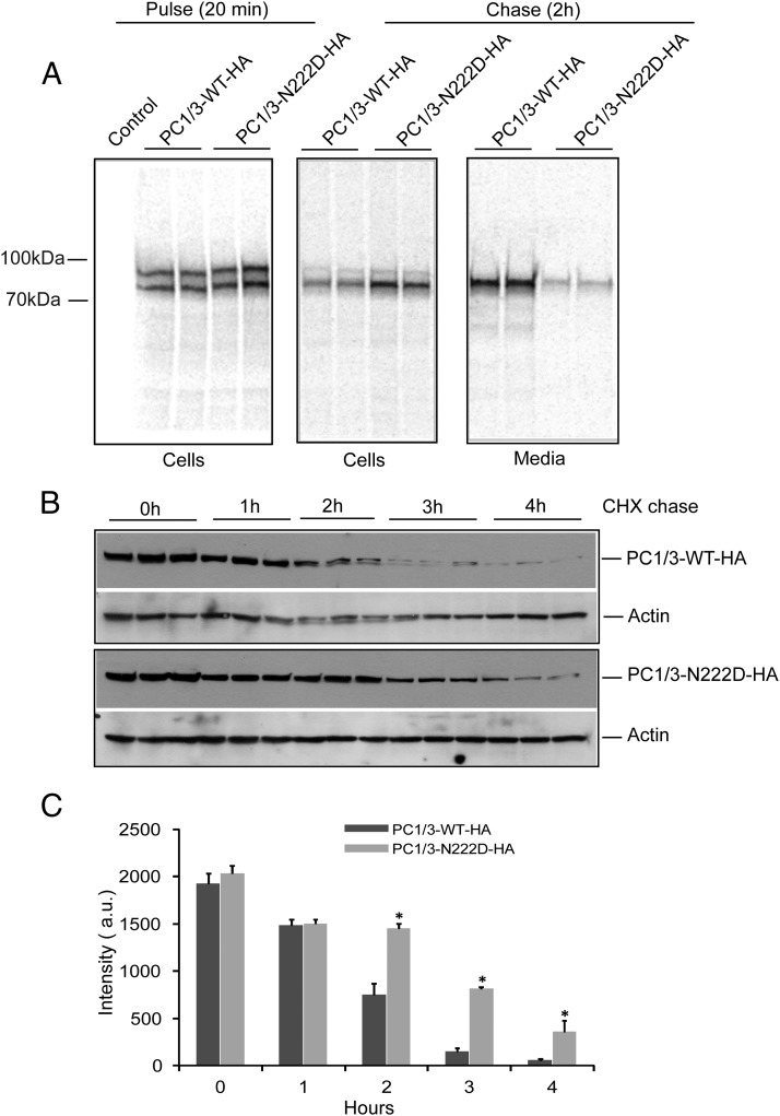
Mutations in the PCSK1 gene encoding prohormone convertase 1/3 (PC1/3) are strongly associated with obesity in humans. The PC1/3(N222D) mutant mouse thus far represents the only mouse model that mimics the PC1/3 obesity phenotype in humans. The present investigation addresses the cell biology of the N222D mutation. Metabolic labeling experiments reveal a clear defect in the kinetics of insulin biosynthesis in islets from PC1/3(N222D) mutant mice, resulting in an increase in both proinsulin and its processing intermediates, predominantly lacking cleavage at the Arg-Arg site. Although the mutant PC1/3 zymogen is correctly processed to the 87-kDa form, pulse-chase immunoprecipitation experiments, labeling, and immunohistochemical experiments using uncleavable variants all demonstrate that the PC1/3-N222D protein is largely mislocalized compared with similar wild-type (WT) constructs, being predominantly retained in the endoplasmic reticulum. The PC1/3-N222D mutant also undergoes more efficient degradation via the ubiquitin-proteasome system than the WT enzyme. Lastly, the mutant PC1/3-N222D protein coimmunoprecipitates with WT PC1/3 and exerts a modest effect on intracellular retention of the WT enzyme. These profound alterations in the cell biology of PC1/3-N222D are likely to contribute to the defective insulin biosynthetic events observed in the mutant mice and may be relevant to the dramatic contributions of polymorphisms in this gene to human obesity.


Figures

Figure 1.
Figure 1.
The PC1/3N222D mutation affects insulin biosynthesis in vivo. A, Schematic showing proinsulin processing. Proinsulin intermediate 1 is cleaved at the C-A junction, and proinsulin intermediate 2 is cleaved at the B-C junction. B, One hundred pancreatic islets were preincubated in DMEM containing 25.5mM glucose for 30 minutes. After being briefly washed twice with Cys/Met-deficient DMEM medium, the islets were pulse labeled with 35S-Cys/Met in the same medium for 20 minutes. The labeled islets were split in half: 1 was lysed immediately, and the other was chased in RPMI 1640 for 4 hours. Newly synthesized proinsulin, conversion intermediates, and insulin were recovered by immunoprecipitation with antiinsulin and analyzed by Tris-tricine-urea-SDS-PAGE under nonreducing conditions. C, Islets were labeled for 20 minutes and chased in RPMI 1640 medium for times indicated. The islets were lysed. The lysates and chase media were combined and immunoprecipitated with antiinsulin followed by Tris-tricine-urea-SDS-PAGE under reducing conditions.
Figure 2.
Figure 2.
The N222D variant of PC1 is localized to the ER, whereas the WT protein is present in the Golgi and in the tips of endocrine cells. A, Schematic representation of PC1/3 showing the prodomain, catalytic domain, P domain, C-terminal domain, mutations incorporated, and the insertion positions of tags. An asterisk represents the stop codon. B, Rin5f cells were transiently transfected with PC1/3-WT-HA and PC1/3-N222D-HA constructs. Cells were fixed with 4% paraformaldehyde and processed as described (19). Colocalization was determined by immunostaining using a 1:100 dilution of monoclonal antibody to HA, a 1:100 dilution of sheep polyclonal antibody anti-TGN38, or a 1:50 dilution of goat polyclonal anti-calreticulin; these were followed by incubation with a 1:250 dilution of Cy2 (green)- or Cy3 (red)-conjugated antimouse immunoglobulin G, Cy2-conjugated antigoat, and Cy3-conjugated antisheep, respectively. C, In order to confirm that differences are due to the N222D mutation, Neuro2a cells were transiently transfected with PC1/3 constructs lacking the mutations that block carboxy-terminal processing. PC1/3-WT-Flag and PC1/3-N222D (untagged) were transfected and were treated as in B, except that PC1/3 was imaged using a rabbit polyclonal anti-PC1/3 antibody (2B5) together with either Cy2- or Cy3-conjugated antirabbit IgG. Scale bar, 10 μm.
Figure 3.
Figure 3.
PC1/3 WT and the PC1/3 N222D variant interact: coimmunoprecipitation. A, Neuro2a cells expressing PC1/3-WT-HA alone or coexpressing either PC1/3-WT-Flag or PC1/3-N222D-Flag were cultured overnight; an unrelated HA-BACE1-WT protein was expressed similarly. Cell lysates were subjected to immunoprecipitation with anti-HA monoclonal antibody and analyzed by tag immunoblotting as indicated. B and C, Neuro2a cells were subjected to transfections in triplicate with plasmids as indicated. Secretion was analyzed by culturing cells overnight in the presence of 0.1 mg/mL aprotinin. Conditioned media and cells were harvested and analyzed by immunoblotting with AAT1 and 2B5 antisera to detect AAT1 and total PC1/3, respectively; or anti-HA antibodies to detect only WT PC1/3. D, Quantification of expression and secretion of WT PC1/3 (HA-tag only) in the presence of N222D PC1/3; WT PC1/3 was normalized to 100%. Data are expressed as the mean ± SD, n = 3. P < .125 for cells (top panel) and P < .01 for media (bottom panel) using an unpaired Student's t test. CO-IP, co-immunoprecipitation; IP, immunoprecipitation.
Figure 4.
Figure 4.
Increased degradation of PC1/3 N222D via the ubiquitination/proteasome system and increased intracellular retention in the presence of MG132. A, Neuro2a cells and HEK cells were transiently transfected with PC1/3-WT-HA or PC1/3-N222D-HA alone or coexpressed with Flag-tagged ubiquitin and cultured in the presence of 5μM lactacystin overnight. Cells were harvested, lysed, and immunoblotted with anti-HA antiserum to detect the immunoprecipitated proteins, and anti-Flag-HRP was then used to detect the ubiquitinated PC1/3. Upper panels show IP, and the lower panel shows ubiquitinated proteins. B, Neuro2a cells were transfected with PC1/3-WT-HA or PC1/3-N222D-HA in triplicate and cultured in the presence of 5μM of the proteasomal inhibitor MG132 for 12 hours. Cells were harvested, lysed, and analyzed for the expression of PC1/3 using an anti-HA antibody. An actin blot was carried out as a loading control. C, Quantification of PC1/3 vs actin expression using GraphPad Prism; data are given as the mean + SD, n = 3. *, P < .05 using an unpaired Student's t test. IP, immunoprecipitation; WB, Western blot.
Figure 5.
Figure 5.
The PC1/3 N222D protein exhibits defective secretion and a longer intracellular half-life than WT PC1/3. A, Pulse-chase assay to assess the biosynthesis of the WT and N222D variant of PC1/3. Neuro2a cells transiently transfected PC1/3-WT-HA and PC1/3-N222D-HA were metabolically labeled with 35S-Cys/Met and chased for 2 hours in medium containing cold methionine. Media were harvested, and cells were harvested, lysed, and immunoprecipitated using anti-2B5 antibody to recover PC1/3 and analyzed as described in Materials and Methods. B, CHX experiment revealing differential degradation profiles of WT PC1/3 and PC1/3-N222D. Transiently transfected PC1/3-WT-HA and PC1/3-N222D-HA constructs were cultured in plating media overnight. Cells were chased in plating media containing 0.1 mg/mL CHX for the indicated time points and analyzed by immunoblotting using anti-HA antibody to detect PC1/3. C, Quantification of B was carried out with ImageJ software, and the data were analyzed using GraphPad Prism, as indicated in the Materials and Methods. (*, P < .05; n = 3; mean ± SD).

Comment in

Similar articles

Cited by

References

    1. Steiner DF. On the discovery of precursor processing. Methods Mol Biol. 2011;768:3–11 - PubMed
    1. Zhou Y, Lindberg I. Enzymatic properties of carboxyl-terminally truncated prohormone convertase 1 (PC1/SPC3) and evidence for autocatalytic conversion. J Biol Chem. 1994;269:18408–18413 - PubMed
    1. Milgram SL, Mains RE. Differential effects of temperature blockade on the proteolytic processing of three secretory granule-associated proteins. J Cell Sci. 1994;107:737–745 - PubMed
    1. Benjannet S, Rondeau N, Paquet L, et al. Comparative biosynthesis, covalent post-translational modifications and efficiency of prosegment cleavage of the prohormone convertases PC1 and PC2: glycosylation, sulphation and identification of the intracellular site of prosegment cleavage of PC1 and PC2. Biochem J. 1993;294(pt 3):735–743 - PMC - PubMed
    1. Coates LC, Birch NP. Posttranslational maturation of the prohormone convertase sPC3 in vitro. J Neurochem. 1997;68:828–836 - PubMed

Publication types

MeSH terms