. 2014 May 14;34(20):6736-45.
doi: 10.1523/JNEUROSCI.5075-13.2014.

α-Melanocyte stimulating hormone prevents GABAergic neuronal loss and improves cognitive function in Alzheimer's disease

Affiliations

α-Melanocyte stimulating hormone prevents GABAergic neuronal loss and improves cognitive function in Alzheimer's disease

Keran Ma et al. J Neurosci. .

Abstract

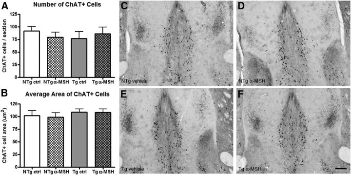
In Alzheimer's disease (AD), appropriate excitatory-inhibitory balance required for memory formation is impaired. Our objective was to elucidate deficits in the inhibitory GABAergic system in the TgCRND8 mouse model of AD to establish a link between GABAergic dysfunction and cognitive function. We sought to determine whether the neuroprotective peptide α-melanocyte stimulating hormone (α-MSH) attenuates GABAergic loss and thus improves cognition. TgCRND8 mice with established β-amyloid peptide pathology and nontransgenic littermates were treated with either α-MSH or vehicle via daily intraperitoneal injections for 28 d. TgCRND8 mice exhibited spatial memory deficits and altered anxiety that were rescued after α-MSH treatment. The expression of GABAergic marker glutamic acid decarboxylase 67 (GAD67) and the number of GABAergic GAD67+ interneurons expressing neuropeptide Y and somatostatin are reduced in the hippocampus in vehicle-treated TgCRND8 mice. In the septohippocampal pathway, GABAergic deficits are observed before cholinergic deficits, suggesting that GABAergic loss may underlie behavior deficits in vehicle-treated TgCRND8 mice. α-MSH preserves GAD67 expression and prevents loss of the somatostatin-expressing subtype of GABAergic GAD67+ inhibitory interneurons. Without decreasing β-amyloid peptide load in the brain, α-MSH improves spatial memory in TgCRND8 mice and prevents alterations in anxiety. α-MSH modulated the excitatory-inhibitory balance in the brain by restoring GABAergic inhibition and, as a result, improved cognition in TgCRND8 mice.

Keywords: Alzheimer's disease; GABAergic system; cognitive function; somatostatin; α-melanocyte stimulating hormone.


Figures

Figure 1.
Figure 1.
Effects of α-MSH on behavior in TgCRND8 mice. Spontaneous alternation analysis of the Y-maze was used to assess the spatial memory of mice. A, The change in percentage alternation shows a significant α-MSH treatment–genotype interaction (p = 0.036). B, At 20 weeks of age, TgCRND8 mice did not exhibit deficits in spatial memory. C, However, at 24 weeks of age, TgCRND8 mice showed a significant decrease in percentage alternation. A, C, α-MSH treatment improved spatial memory in TgCRND8 mice (p < 0.001) with no effect on non-Tg littermates (p = 0.786). D, In the open field test, there is a significant interaction between α-MSH treatment and genotype (p = 0.015). E, F, TgCRND8 vehicle-treated mice showed a significant decrease in anxiety from 20 to 24 weeks of age and α-MSH treatment prevented this change (p = 0.008). Locomotion of mice was also measured in the open field test. G, There is no significant interaction between α-MSH treatment and genotype in the total distance traveled (p = 0.887). H, I, Both vehicle-treated and α-MSH-treated TgCRND8 mice are more hyperactive compared with non-Tg mice (p < 0.001). NTg ctrl, Vehicle-treated NTg mice; NTg α-MSH, α-MSH-treated NTg mice; Tg ctrl, vehicle-treated Tg mice; Tg α-MSH, α-MSH-treated Tg mice. Data represent mean ± SEM, n = 10–23 per group, *p < 0.05.
Figure 2.
Figure 2.
Aβ accumulation as a function of α-MSH treatment. A–D, α-MSH had no effect on insoluble or soluble levels of Aβ40 (A, B) or Aβ42 (C, D) in the hippocampus and the cortex. E, F, The number of Aβ plaques (p = 0.72; E) or area covered by plaques (p = 0.66; F) in the hippocampus are also not affected by α-MSH. G, H, Representative photomicrographs of Aβ plaque staining in vehicle-treated (G) and α-MSH TgCRND8-treated (H) mice. Data represent mean ± SEM, n = 5–6 per group. Scale bar, 300 μm.
Figure 3.
Figure 3.
GABAergic marker expression in the hippocampus. A, B, GAD67 mRNA (A) and protein levels (B) are significantly decreased in TgCRND8 mice compared with non-Tg littermates (p = 0.044 and p = 0.001 respectively). C, GAD65 protein levels are not changed with genotype or α-MSH treatment. α-MSH treatment rescues the GAD67 deficits in TgCRND8 mice in both mRNA (p = 0.008) and protein levels (p = 0.009). Data represent mean ± SEM, n = 6–7 per group, *p < 0.05.
Figure 4.
Figure 4.
Effect of α-MSH treatment on GABAergic GAD67+ interneurons in the hippocampus. A, The low number of GAD67+ cells in the CA1 region of the hippocampus in TgCRND8 mice (p = 0.018; A) increases with α-MSH treatment (p = 0.016). B, C, Neither genotype nor α-MSH treatment changed the number of GAD67+ cells in the CA3 region (B) or the hilus (C) of the hippocampus. D–G, Representative photomicrographs of the GAD67+ GABAergic interneurons in the CA1 region of the hippocampus in NTg vehicle-treated (D), NTg α-MSH-treated (E), Tg vehicle-treated (F), and Tg α-MSH-treated (G) mice. Data represent mean ± SEM, n = 6–7 per group, *p < 0.05. Scale bar, 100 μm.
Figure 5.
Figure 5.
ChAT+ cholinergic phenotype in the medial septum. A, B, TgCRND8 mice do not show a decrease in the number or area of ChAT+ cells in the medial septum (p = 0.38 and p = 0.58 respectively). α-MSH treatment does not alter the numbers or area of ChAT+ cells in either TgCRND8 (number, p = 0.57; area, p = 0.95) or NTg mice (number, p = 0.46; area, p = 0.77). C–F, Representative photomicrographs of the ChAT+ cholinergic cells in the medial septum in NTg vehicle-treated (C), NTg α-MSH-treated (D), Tg vehicle-treated (E), and Tg α-MSH-treated (F) mice. Data represent mean ± SEM, n = 6 per group. Scale bar, 100 μm.
Figure 6.
Figure 6.
Effect of α-MSH treatment on GABAergic neuronal subtypes in the hippocampus. GABAergic neurons can be divided into subtypes according to the neuropeptide expressed. A–C, In the hippocampus, mRNA expression of NPY (A) and SST (B) is significantly decreased in TgCRND8 mice (p = 0.01 and p = 0.038 respectively), but not CCK (C). α-MSH treatment rescued SST mRNA levels (B) but does not affect NPY (A) or CCK (C) expression. Data represent mean ± SEM, n = 6–7 per group, *p < 0.05.
Figure 7.
Figure 7.
Effect of α-MSH treatment on SST+ cells in the hippocampus. A, SST+ cells in the stratum oriens of the CA1 is significantly decreased in TgCRND8 mice and rescued by α-MSH treatment. B, C, Neither genotype nor α-MSH treatment altered the number of SST+ cells in the CA3 region (B) or the hilus (C) of the hippocampus. D–G, Representative photomicrographs of the SST+ cells in the CA1 region of the hippocampus in NTg vehicle-treated (D), NTg α-MSH-treated (E), Tg vehicle-treated (F), and Tg α-MSH-treated (G) mice. H, SST+ cells colocalize with GAD67+ GABAergic interneurons in the CA1 stratum oriens of the hippocampus. Data represent mean ± SEM, n = 6–11 per group, *p < 0.05. Scale bars: SST IHC, 100 μm; GAD67/SST colocalization, 25 μm.

Similar articles

Cited by

References

    1. Alom J, Galard R, Catalan R, Castellanos JM, Schwartz S, Tolosa E. Cerebrospinal fluid neuropeptide Y in Alzheimer's disease. Eur Neurol. 1990;30:207–210. doi: 10.1159/000117347. - DOI - PubMed
    1. Andrews-Zwilling Y, Bien-Ly N, Xu Q, Li G, Bernardo A, Yoon SY, Zwilling D, Yan TX, Chen L, Huang Y. Apolipoprotein E4 causes age- and Tau-dependent impairment of GABAergic interneurons, leading to learning and memory deficits in mice. J Neurosci. 2010;30:13707–13717. doi: 10.1523/JNEUROSCI.4040-10.2010. - DOI - PMC - PubMed
    1. Arai H, Moroji T, Kosaka K, Iizuka R. Extrahypophyseal distribution of alpha-melanocyte stimulating hormone (alpha-MSH)-like immunoreactivity in postmortem brains from normal subjects and Alzheimer-type dementia patients. Brain Res. 1986;377:305–310. doi: 10.1016/0006-8993(86)90873-5. - DOI - PubMed
    1. Bellucci A, Luccarini I, Scali C, Prosperi C, Giovannini MG, Pepeu G, Casamenti F. Cholinergic dysfunction, neuronal damage and axonal loss in TgCRND8 mice. Neurobiol Dis. 2006;23:260–272. doi: 10.1016/j.nbd.2006.03.012. - DOI - PubMed
    1. Buzsáki G. Theta oscillations in the hippocampus. Neuron. 2002;33:325–340. doi: 10.1016/S0896-6273(02)00586-X. - DOI - PubMed

Publication types

MeSH terms