

Chikungunya viruses that escape monoclonal antibody therapy are clinically attenuated, stable, and not purified in mosquitoes
- PMID: 24829346
- PMCID: PMC4135940
- DOI: 10.1128/JVI.01032-14
Chikungunya viruses that escape monoclonal antibody therapy are clinically attenuated, stable, and not purified in mosquitoes
Abstract
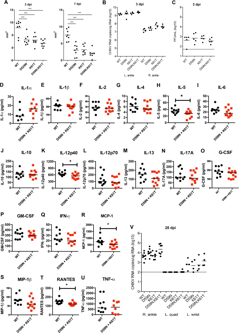
Chikungunya virus (CHIKV) is a reemerging mosquito-transmitted alphavirus that causes epidemics of debilitating polyarthritis in humans. A prior study identified two anti-CHIKV monoclonal antibodies ([MAbs] CHK-152 and CHK-166) against the E2 and E1 structural proteins, which had therapeutic efficacy in immunocompetent and immunocompromised mice. Combination MAb therapy was required as administration of a single MAb resulted in the rapid selection of neutralization escape variants and treatment failure in mice. Here, we initially evaluated the efficacy of combination MAb therapy in a nonhuman primate model of CHIKV infection. Treatment of rhesus macaques with CHK-152 and CHK-166 reduced viral spread and infection in distant tissue sites and also neutralized reservoirs of infectious virus. Escape viruses were not detected in the residual viral RNA present in tissues and organs of rhesus macaques. To evaluate the possible significance of MAb resistance, we engineered neutralization escape variant viruses (E1-K61T, E2-D59N, and the double mutant E1-K61T E2-D59N) that conferred resistance to CHK-152 and CHK-166 and tested them for fitness in mosquito cells, mammalian cells, mice, and Aedes albopictus mosquitoes. In both cell culture and mosquitoes, the mutant viruses grew equivalently and did not revert to wild-type (WT) sequence. All escape variants showed evidence of mild clinical attenuation, with decreased musculoskeletal disease at early times after infection in WT mice and a prolonged survival time in immunocompromised Ifnar1(-/-) mice. Unexpectedly, this was not associated with decreased infectivity, and consensus sequencing from tissues revealed no evidence of reversion or compensatory mutations. Competition studies with CHIKV WT also revealed no fitness compromise of the double mutant (E1-K61T E2-D59N) neutralization escape variant in WT mice. Collectively, our study suggests that neutralization escape viruses selected during combination MAb therapy with CHK-152 plus CHK-166 retain fitness, cause less severe clinical disease, and likely would not be purified during the enzootic cycle.
Importance: Chikungunya virus (CHIKV) causes explosive epidemics of acute and chronic arthritis in humans in Africa, the Indian subcontinent, and Southeast Asia and recently has spread to the New World. As there are no approved vaccines or therapies for human use, the possibility of CHIKV-induced debilitating disease is high in many parts of the world. To this end, our laboratory recently generated a combination monoclonal antibody therapy that aborted lethal and arthritogenic disease in wild-type and immunocompromised mice when administered as a single dose several days after infection. In this study, we show the efficacy of the antibody combination in nonhuman primates and also evaluate the significance of possible neutralization escape mutations in mosquito and mammalian cells, mice, and Aedes albopictus vector mosquitoes. Our experiments show that escape viruses from combination antibody therapy cause less severe CHIKV clinical disease, retain fitness, and likely would not be purified by mosquito vectors.
Copyright © 2014, American Society for Microbiology. All Rights Reserved.
Figures
Similar articles
-
Development of a highly protective combination monoclonal antibody therapy against Chikungunya virus.PLoS Pathog. 2013;9(4):e1003312. doi: 10.1371/journal.ppat.1003312. Epub 2013 Apr 18. PLoS Pathog. 2013. PMID: 23637602 Free PMC article.
-
Chikungunya Virus Fidelity Variants Exhibit Differential Attenuation and Population Diversity in Cell Culture and Adult Mice.J Virol. 2019 Jan 17;93(3):e01606-18. doi: 10.1128/JVI.01606-18. Print 2019 Feb 1. J Virol. 2019. PMID: 30429348 Free PMC article.
-
Emerging Chikungunya Virus Variants at the E1-E1 Interglycoprotein Spike Interface Impact Virus Attachment and Inflammation.J Virol. 2022 Feb 23;96(4):e0158621. doi: 10.1128/JVI.01586-21. Epub 2021 Dec 22. J Virol. 2022. PMID: 34935436 Free PMC article.
-
Chikungunya virus: evolution and genetic determinants of emergence.Curr Opin Virol. 2011 Oct;1(4):310-7. doi: 10.1016/j.coviro.2011.07.004. Curr Opin Virol. 2011. PMID: 21966353 Free PMC article. Review.
-
Chikungunya virus and its mosquito vectors.Vector Borne Zoonotic Dis. 2015 Apr;15(4):231-40. doi: 10.1089/vbz.2014.1745. Epub 2015 Feb 12. Vector Borne Zoonotic Dis. 2015. PMID: 25674945 Review.
Cited by
-
Therapeutic administration of a recombinant human monoclonal antibody reduces the severity of chikungunya virus disease in rhesus macaques.PLoS Negl Trop Dis. 2017 Jun 19;11(6):e0005637. doi: 10.1371/journal.pntd.0005637. eCollection 2017 Jun. PLoS Negl Trop Dis. 2017. PMID: 28628616 Free PMC article.
-
Optimal therapeutic activity of monoclonal antibodies against chikungunya virus requires Fc-FcγR interaction on monocytes.Sci Immunol. 2019 Feb 22;4(32):eaav5062. doi: 10.1126/sciimmunol.aav5062. Sci Immunol. 2019. PMID: 30796092 Free PMC article.
-
Vertebrate Reservoirs of Arboviruses: Myth, Synonym of Amplifier, or Reality?Viruses. 2017 Jul 13;9(7):185. doi: 10.3390/v9070185. Viruses. 2017. PMID: 28703771 Free PMC article. Review.
-
Evolution of antiviral resistance captures a transient interdomain functional interaction between chikungunya virus envelope glycoproteins.bioRxiv [Preprint]. 2024 Nov 11:2024.11.11.623010. doi: 10.1101/2024.11.11.623010. bioRxiv. 2024. PMID: 39605706 Free PMC article. Preprint.
-
Tropism of the Chikungunya Virus.Viruses. 2019 Feb 20;11(2):175. doi: 10.3390/v11020175. Viruses. 2019. PMID: 30791607 Free PMC article. Review.
References
Publication types
MeSH terms
Substances
Grants and funding
LinkOut - more resources
Full Text Sources
Other Literature Sources
Medical
Research Materials
