. 2014 Jul 15;25(14):2171-80.
doi: 10.1091/mbc.E14-03-0842. Epub 2014 May 14.

Dynactin integrity depends upon direct binding of dynamitin to Arp1

Affiliations

Dynactin integrity depends upon direct binding of dynamitin to Arp1

Frances Ka Yan Cheong et al. Mol Biol Cell. .

Abstract

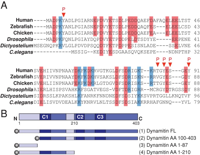
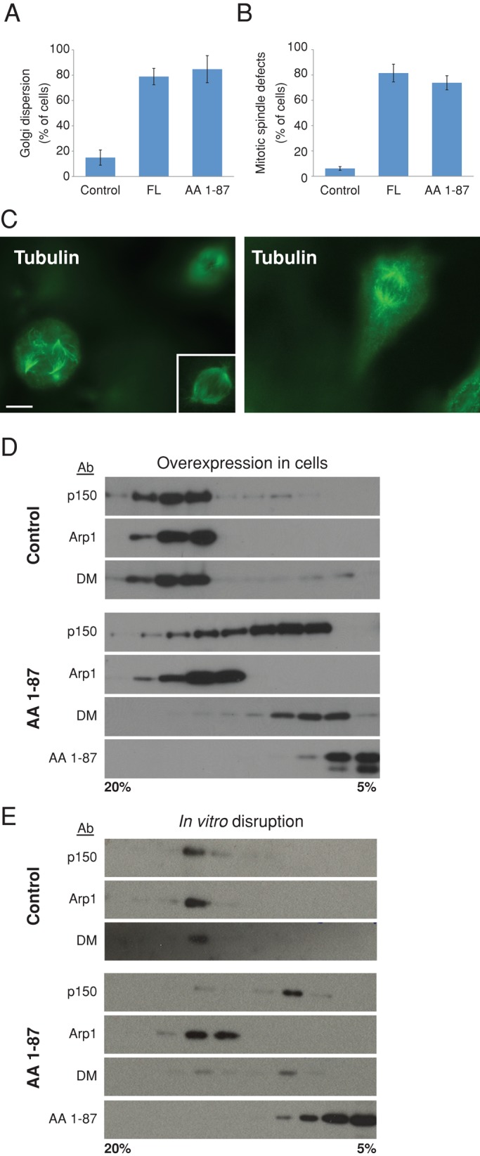
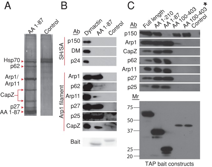
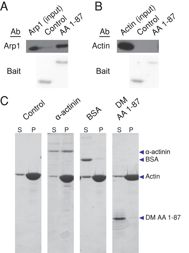
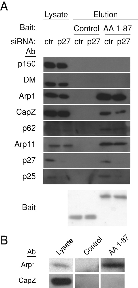
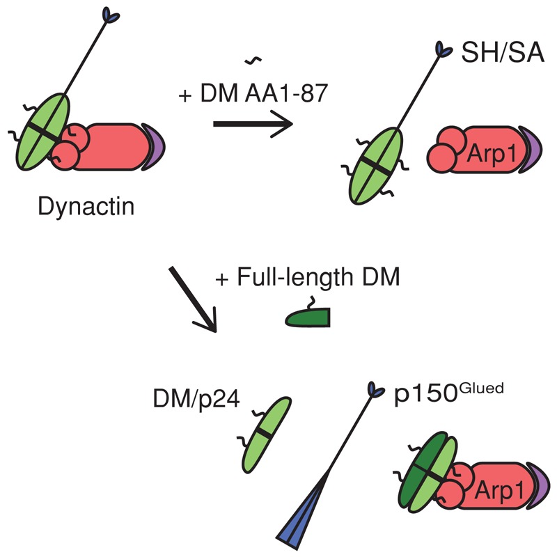
Dynactin is a multiprotein complex that works with cytoplasmic dynein and other motors to support a wide range of cell functions. It serves as an adaptor that binds both dynein and cargoes and enhances single-motor processivity. The dynactin subunit dynamitin (also known as p50) is believed to be integral to dynactin structure because free dynamitin displaces the dynein-binding p150(Glued) subunit from the cargo-binding Arp1 filament. We show here that the intrinsically disordered dynamitin N-terminus binds to Arp1 directly. When expressed in cells, dynamitin amino acids (AA) 1-87 causes complete release of endogenous dynamitin, p150, and p24 from dynactin, leaving behind Arp1 filaments carrying the remaining dynactin subunits (CapZ, p62, Arp11, p27, and p25). Tandem-affinity purification-tagged dynamitin AA 1-87 binds the Arp filament specifically, and binding studies with purified native Arp1 reveal that this fragment binds Arp1 directly. Neither CapZ nor the p27/p25 dimer contributes to interactions between dynamitin and the Arp filament. This work demonstrates for the first time that Arp1 can directly bind any protein besides another Arp and provides important new insight into the underpinnings of dynactin structure.


Figures

FIGURE 1:
FIGURE 1:
Dynamitin primary structure. (A) Sequence alignment of the dynamitin N-terminus of selected nonfungal species. Acidic residues are shaded red, basic residues are shaded blue, and identified in vivo phosphorylation sites (www.phosphosite.org) are indicated by red arrowheads. (B) Top, the dynamitin primary sequence, indicating predicted structural motifs, including unstructured motifs (light blue; AA 1–105 and AA 186–213) and multicoil motifs C1 (AA 105–135), C2 (AA 219–251), and C3 (281–308). Bottom, cartoons of the dynamitin constructs used in this study. Each construct was tagged (shaded circles) at the N-terminus with TAP, 6X-His, myc, or mCherry as specified in the Results. Note that the fragment that is the focus of this work (chicken dynamitin AA 1–87) is different from the previously published chicken AA 1–78 fragment (Maier et al., 2008; Valetti et al., 1999).
FIGURE 2:
FIGURE 2:
Effect of dynamitin AA 1–87 on dynactin structure and function in vitro and in vivo. (A, B) cDNAs encoding myc-tagged proteins were introduced into Cos-7 cells ∼24 h before fixation and staining for giantin to visualize the Golgi complex or tubulin to visualize mitotic spindles. Overexpressing cell populations were scored for the presence of a dispersed Golgi complex (A) or deranged spindle (B; mean ± SD; three experiments; 700 < n < 800 cells per condition for Golgi and n = 400 per condition for spindles). The results shown here were obtained using myc-tagged proteins, but similar results were obtained using mCherry-tagged dynamitin species. Myc-tagged dynactin p62 AA 370–467 and monomeric red fluorescent protein (mRFP) were used as controls in A, and CMV-β gal was used as a control in B. (C) Left, representative image of a cell expressing myc-tagged AA 1–87, stained for tubulin. The nonexpressing cell at the upper right is in a different focal plane, and the inset shows this cell's spindle in focus. (See Supplemental Figure S1 for a merged image showing myc staining.) Right, A control cell expressing CMV-β gal, stained for tubulin. Scale bar, 5 μm. (D) cDNA encoding TAP-tagged AA 1–87 (or buffer as a control) was electroporated into Cos-7 cells. After 48 h, detergent lysates were subjected to velocity sedimentation into a 5–20% sucrose gradient. Gradient fractions were analyzed by immunoblotting to detect the dynactin subunit p150Glued, Arp1, or dynamitin (DM). Dynamitin AA 1–87 was detected using an antibody to TAP. Similar results were obtained with myc- or mCherry-tagged AA 1–87. (E) Purified bovine dynactin (10 μg) was mixed with 100× molar excess of recombinant (6X-His) dynamitin AA 1–87 and subjected to velocity sedimentation as in D. AA 1–87 was detected using an antibody to the Xpress tag.
FIGURE 3:
FIGURE 3:
Isolation of binding partners for dynamitin N- and C-terminal fragments. (A) cDNAs encoding TAP-tagged AA 1–87 or a control (TAP-Mef2a, provided by Stratagene) were electroporated into Cos-7 cells, then lysates were prepared and subjected to tandem affinity purification on streptavidin and calmodulin resins as described in Materials and Methods. The final eluates were evaluated on a silver-stained SDS–polyacrylamide gel. Dynactin subunits (and Hsp70, the only other major component of the AA 1–87 eluate) are indicated with arrowheads. (B) Beads bearing purified 6X-His-AA 1–87 or a control (6X-His-TrbB) were mixed with purified bovine dynactin, and proteins were eluted using imidazole (see Materials and Methods). Samples were analyzed by immunoblotting. Arp1 filament and shoulder/arm (Sh/SA) components are indicated. p62 and Arp11 are very minor components that can behave erratically on blots, explaining the relatively weak signal. The “bait” protein present in each sample (6X-His-AA 1–87 or 6X-His TrbB) was detected by Ponceau S staining of the PVDF membrane. (C) Top, lysates prepared from cells expressing TAP-tagged dynamitin fragments or an untransfected control were incubated with streptavidin beads. Binding partners were eluted and analyzed by immunoblotting for the proteins indicated. For the sample labeled AA 100–403*, the lysate overexpressing TAP AA 100–403 was supplemented with recombinant 6X-His-AA 1–87 to release the shoulder/arm complex from the Arp1 filament before addition of beads. Bottom, TAP-tagged “bait” proteins present in the bead eluates were detected by immunoblotting (for calmodulin-binding protein).
FIGURE 4:
FIGURE 4:
Analysis of dynamitin AA 1–87 binding to Arp1 and actin. (A, B) Arp1 isolated from bovine dynactin by KI treatment and gel filtration (A; Bingham and Schroer, 1999) or G-actin (B) was dialyzed into G-buffer for 1 h and then mixed with Talon beads bearing purified 6X-His-AA 1–87 or a control (6X-His-Fis1ΔTM). Samples were then analyzed by immunoblotting to detect Arp1 (2% of input and 2.5% of beads) or actin (2% of input and beads). Bait proteins were detected on the PVDF membrane by Ponceau S staining. (C) F-actin (see Materials and Methods) was incubated with buffer alone, α-actinin, BSA, or purified 6X-His-AA 1-87 (DM AA 1–87) for 30 min at room temperature. F-actin and bound proteins were then pelleted, and equal proportions of the supernatants (S) and pellets (P) were evaluated by SDS–PAGE, followed by Coomassie blue staining.
FIGURE 5:
FIGURE 5:
Analysis of the contributions of p27/p25 and CapZ to dynamitin–Arp1 binding. (A) Lysates of Cos-7 cells electroporated with p27 or control (ctr) siRNAs were incubated with 6X-His-AA 1–87 or a control (Fis1∆TM) before addition of Talon beads. Dynactin subunits in the eluates were detected by immunoblotting (1% of lysate and 5% of eluates). Bait proteins were detected by Ponceau S staining of the PVDF membrane. (B) Pools of cytosolic proteins (“lysate” lane) containing dynactin (top: 20S fraction) or free CapZ (bottom; 4–5S fraction) were separated by sucrose gradient sedimentation and then mixed with beads bearing 6X-His-AA 1-87 or a control (6X His-TrbB). The 4–5S fractions (bottom left) and the bead eluates (bottom middle and right) were immunoblotted for CapZ. Binding of Arp1 (in the 20S fraction; top left) to control or AA 1–87 beads was assayed in parallel (top middle and right).
FIGURE 6:
FIGURE 6:
Proposed model of dynamitin interactions with other dynactin components. The unstructured dynamitin N-terminus (AA 1–87; black squiggle) binds directly to Arp1 (red) to anchor the shoulder and arm (SH/SA) structure to the Arp filament. The remainder of dynamitin (light green) is engaged in interactions with other dynamitin protomers and with p24 (thick black line) Top, excess AA 1–87 (DM 1–87) displaces the entire shoulder and arm structure from the Arp filament (purple depicts the pointed-end complex: p62, Arp11, p27, p25). Bottom, exogenous full-length dynamitin (DM, dark green) triggers subunit release by interacting with other shoulder/arm components and remodeling their interactions. This leads to displacement of a dynamitin/p24 (2:1) complex (light green; the thick black line bisecting the oval represents one p24 protomer) and the p150Glued dimer (blue). Two exogenous dynamitin protomers are exchanged for two endogenous protomers (Melkonian et al., 2007).

References

    1. Amaro IA, Costanzo M, Boone C, Huffaker TC. The Saccharomyces cerevisiae homolog of p24 is essential for maintaining the association of p150Glued with the dynactin complex. Genetics. 2008;178:703–709. - PMC - PubMed
    1. Bingham JB, Schroer TA. Self-regulated polymerization of the actin-related protein. Arp1 Curr Biol. 1999;9:223–226. - PubMed
    1. Brown CL, Maier KC, Stauber T, Ginkel LM, Wordeman L, Vernos I, Schroer TA. Kinesin-2 is a motor for late endosomes and lysosomes. Traffic. 2005;6:1114–1124. - PubMed
    1. Burkhardt JK, Echeverri CJ, Nilsson T, Vallee RB. Overexpression of the dynamitin (p50) subunit of the dynactin complex disrupts dynein-dependent maintanance of membrane organelle distribution. J Cell Biol. 1997;139:469–484. - PMC - PubMed
    1. Chen YH, Yang JT. A new approach to the calculation of secondary structures of globular proteins by optical rotatory dispersion and circular dichroism. Biochem Biophys Res Commun. 1971;44:1285–1291. - PubMed

Publication types

LinkOut - more resources