. 2014 Jun 15;192(12):6053-61.
doi: 10.4049/jimmunol.1301481. Epub 2014 May 14.

IL-36 promotes myeloid cell infiltration, activation, and inflammatory activity in skin

Affiliations

IL-36 promotes myeloid cell infiltration, activation, and inflammatory activity in skin

Alexander M Foster et al. J Immunol. .

Abstract

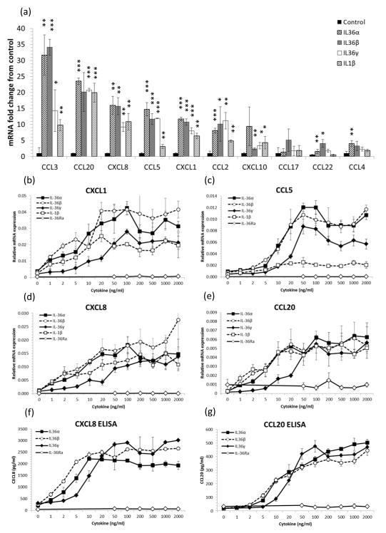
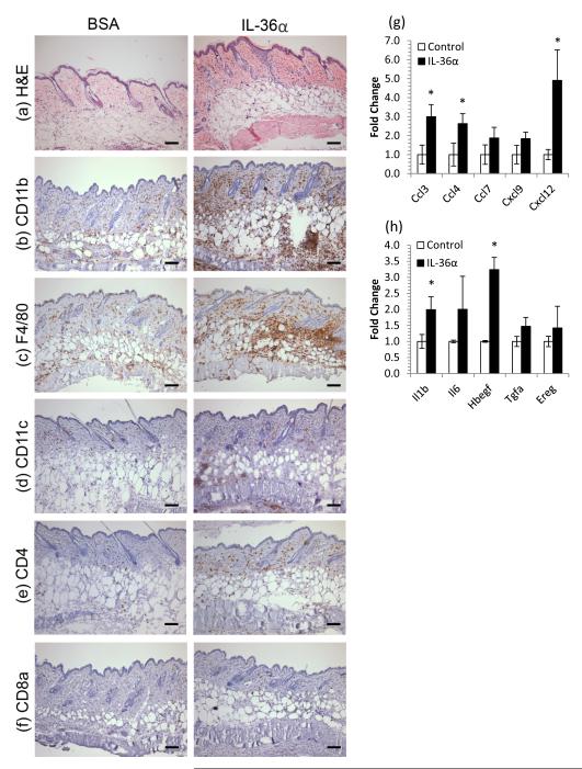
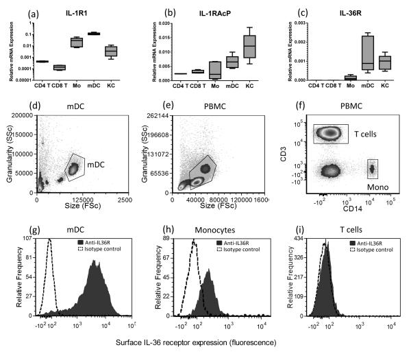
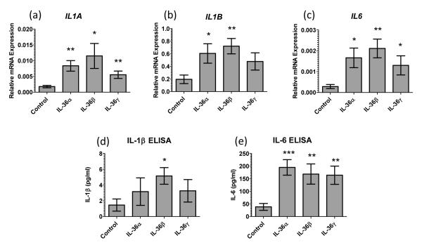
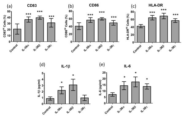
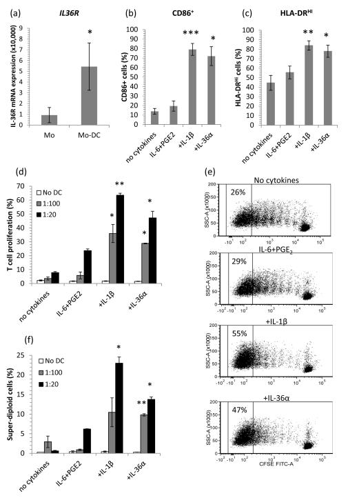
The IL-1 family members IL-36α (IL-1F6), IL-36β (IL-1F8), and IL-36γ (IL-1F9) and the receptor antagonist IL-36Ra (IL-1F5) constitute a novel signaling system that is poorly understood. We now show that these cytokines have profound effects on the skin immune system. Treatment of human keratinocytes with IL-36 cytokines significantly increased the expression of CXCL1, CXCL8, CCL3, CCL5, and CCL20, potent chemotactic agents for activated leukocytes, and IL-36α injected intradermally resulted in chemokine expression, leukocyte infiltration, and acanthosis of mouse skin. Blood monocytes, myeloid dendritic cells (mDC), and monocyte-derived DC (MO-DC) expressed IL-36R and responded to IL-36. In contrast, no direct effects of IL-36 on resting or activated human CD4(+) or CD8(+) T cells, or blood neutrophils, could be demonstrated. Monocytes expressed IL-1A, IL-1B, and IL-6 mRNA and IL-1β and IL-6 protein, and mDC upregulated surface expression of CD83, CD86, and HLA-DR and secretion of IL-1β and IL-6 after treatment with IL-36. Furthermore, IL-36α-treated MO-DC enhanced allogeneic CD4(+) T cell proliferation, demonstrating that IL-36 can stimulate the maturation and function of DC and drive T cell proliferation. These data indicate that IL-36 cytokines actively propagate skin inflammation via the activation of keratinocytes, APC, and, indirectly, T cells.


Figures

Figure 1
Figure 1. IL-36 cytokines induce chemokine expression by keratinocytes
4-day post-confluent normal human keratinocytes (NHK) were stimulated for 24h with recombinant truncated IL-36R ligands or IL-1β. Total RNA was extracted and mRNA transcripts quantified by qRT-PCR relative to the housekeeping gene RPL-P0 and conditioned medium assayed by ELISA. 100ng/ml IL-36α, IL-36β and IL-36γ significantly induced T cell chemokine mRNA expression compared with untreated cells, mean ± S.D. (n=3) (a). IL-36α, IL-36β, IL-36γ and IL-1β but not IL-36Ra dose-dependently induced CXCL1, CCL5, CXCL8 and CCL20 mRNA expression (b-e) and CXCL8 and CCL20 protein secretion (f and g) by keratinocytes. Mean ± S.D. (n=3). Statistical significance indicated by * p<0.05, t** p<0.01 or *** p<0.001, Student’s t-test.
Figure 2
Figure 2. IL-36 induces myeloid cell infiltration of skin concomitant with chemokine and growth factor induction
5μg rmIL-36α or BSA was injected intradermally into CD1 mice every other day for 10 days. Back skin was harvested, snap frozen, processed for RNA and histochemistry. IL-36α treatment lead to acanthosis and an increase in eosinophilic dermal collagen (a) and striking infiltration of granulocytes (CD11b, b), macrophages (F4/80, c), dendritic cells (CD11c, d) CD4+ cells (e) but not CD8+ cells (f). These changes were accompanied by increases in chemokines (g) and cytokines/growth factors (h). Mean ± SEM (n=4 mice). Statistical significance indicated by * p<0.05 (2-tailed t-test). Scale bar = 100μm.
Figure 3
Figure 3. Human APC but not T cells express the IL-36 receptor
Keratinocytes (KC), monocytes and myeloid DC (mDC) express IL-1R1, IL-1RAcP and IL-36R mRNA transcripts, however, CD4+ and CD8+ T cells were found not to express IL-36R as determined by qRT-PCR (a-c, n=4 donors). Flow cytometric analysis reveals that in contrast to mDC (d and g) and monocytes (e, f and h), T cells (e, f and i) did not express surface IL-36R. Filled histogram: anti-IL36R, dotted histogram, isotype control antibody. Flow cytometry gating shown in panels 3d-f; Flow cytometry data are representative of 6 donors.
Figure 4
Figure 4. IL-36 cytokines induce monocyte expression of inflammatory cytokines
Monocytes treated with 100ng/ml IL-36 cytokines for 12h upregulate IL-1A, IL-1B and IL-6, cytokine transcripts (n=9 donors, a-c). Significantly elevated levels of IL-1β and IL-6 were detected in the conditioned culture media after 12h (n=6 donors, e and f). Bars, mean ± SEM. Statistical significance indicated by * p<0.05, ** p<0.01, *** p<0.001 (2-tailed t-test).
Figure 5
Figure 5. IL-36 cytokines facilitate myeloid DC activation and cytokine secretion
Ex vivo blood myeloid DC treated with 100ng/ml IL-36 cytokines for 12h up-regulate CD83 (a), CD86 (b) and HLA-DR (c) expression (FACS, n=6 donors) and secretion of IL-1B (d) and IL-6 (e) (ELISA, n=9). Bars, mean ± SD. Statistical significance indicated by * p<0.05, ** p<0.01, *** p<0.001 (2-tailed t-test).
Figure 6
Figure 6. Monocyte-derived dendritic cells (MO-DC) have enhanced IL-36R expression and IL-36 cytokines facilitate DC maturation, driving T cell proliferation
MO-DC expressed 6-fold more IL-36R mRNA than their monocyte precursors (qRT-PCR, n=6, a). When cultured with 10ng/ml IL-6+0.1μM PGE2 and either 10ng/ml IL-1β or 100ng/ml IL-36α, MODC significantly increased their surface expression of CD86 (b) and HLA-DR (c), indicative of DC maturation (48hr, bars, mean ± SEM, n=8 donors). MO-DC matured with 10ng/ml IL-6 + 0.1μM PGE2 and 10ng/ml TNF-α, 10ng/ml IL-1β or 100ng/ml IL-36α drove allogeneic T cell proliferation as determined by CFSE dye dilution (d and e) or DAPI labeling of DNA (f). No DC (T cells only), 1:100 DC:T cell ratio, 1:20 DC:T cell ratio showing mean ± SEM (n=4 donors). Statistical significance indicated by * p<0.05, ** p<0.01, *** p<0.001 compared with IL-6+PGE2 (2-tailed t-test).

References

    1. Blumberg H, Dinh H, Trueblood ES, Pretorius J, Kugler D, Weng N, Kanaly ST, Towne JE, Willis CR, Kuechle MK, Sims JE, Peschon JJ. Opposing activities of two novel members of the IL-1 ligand family regulate skin inflammation. J Exp Med. 2007;204:2603–2614. - PMC - PubMed
    1. Towne JE, Garka KE, Renshaw BR, Virca GD, Sims JE. Interleukin (IL)-1F6, IL-1F8, and IL-1F9 signal through IL-1Rrp2 and IL-1RAcP to activate the pathway leading to NF-kappaB and MAPKs. J Biol Chem. 2004;279:13677–13688. - PubMed
    1. Magne D, Palmer G, Barton JL, Mezin F, Talabot-Ayer D, Bas S, Duffy T, Noger M, Guerne PA, Nicklin MJ, Gabay C. The new IL-1 family member IL-1F8 stimulates production of inflammatory mediators by synovial fibroblasts and articular chondrocytes. Arthritis Res Ther. 2006;8:R80. - PMC - PubMed
    1. Debets R, Timans JC, Homey B, Zurawski S, Sana TR, Lo S, Wagner J, Edwards G, Clifford T, Menon S, Bazan JF, Kastelein RA. Two novel IL-1 family members, IL-1 delta and IL-1 epsilon, function as an antagonist and agonist of NF-kappa B activation through the orphan IL-1 receptor-related protein 2. J Immunol. 2001;167:1440–1446. - PubMed
    1. Johnston A, Xing X, Guzman AM, Riblett M, Loyd CM, Ward NL, Wohn C, Prens EP, Wang F, Maier LE, Kang S, Voorhees JJ, Elder JT, Gudjonsson JE. IL-1F5, -F6, -F8, and -F9: a novel IL-1 family signaling system that is active in psoriasis and promotes keratinocyte antimicrobial peptide expression. J Immunol. 2011;186:2613–2622. - PMC - PubMed

Publication types

MeSH terms