. 2014 Jun 15;192(12):5906-12.
doi: 10.4049/jimmunol.1302992. Epub 2014 May 14.

Lymphopenia associated with highly virulent H5N1 virus infection due to plasmacytoid dendritic cell-mediated apoptosis of T cells

Affiliations

Lymphopenia associated with highly virulent H5N1 virus infection due to plasmacytoid dendritic cell-mediated apoptosis of T cells

Kobporn Boonnak et al. J Immunol. .

Abstract

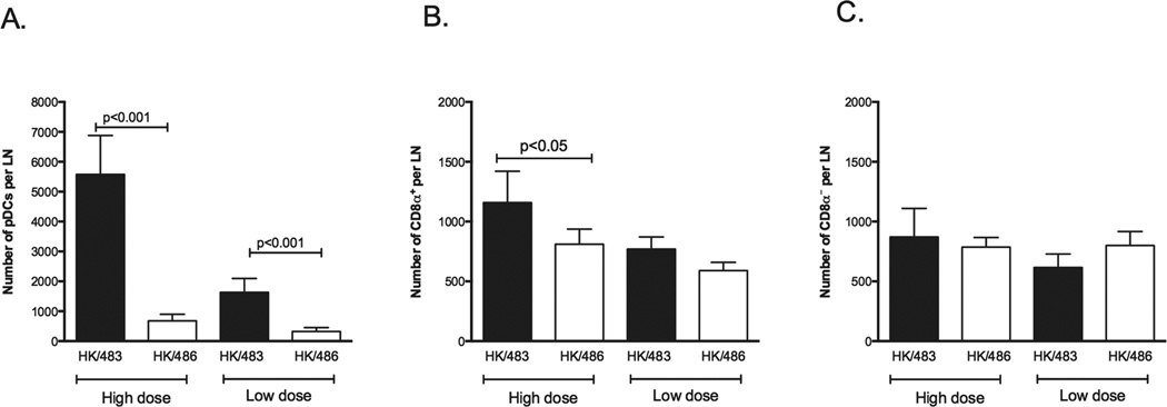
Although lymphopenia is a hallmark of severe infection with highly pathogenic H5N1 and the newly emerged H7N9 influenza viruses in humans, the mechanism(s) by which lethal H5N1 viruses cause lymphopenia in mammalian hosts remains poorly understood. Because influenza-specific T cell responses are initiated in the lung draining lymph nodes (LNs), and lymphocytes subsequently traffic to the lungs or peripheral circulation, we compared the immune responses in the lung draining LNs postinfection with a lethal A/HK/483/97 or nonlethal A/HK/486/97 (H5N1) virus in a mouse model. We found that lethal H5N1, but not nonlethal H5N1, virus infection in mice enhances Fas ligand (FasL) expression on plasmacytoid dendritic cells (pDCs), resulting in apoptosis of influenza-specific CD8(+) T cells via a Fas-FasL-mediated pathway. We also found that pDCs, but not other DC subsets, preferentially accumulate in the lung draining LNs of lethal H5N1 virus-infected mice, and that the induction of FasL expression on pDCs correlates with high levels of IL-12p40 monomer/homodimer in the lung draining LNs. Our data suggest that one of the mechanisms of lymphopenia associated with lethal H5N1 virus infection involves a deleterious role for pDCs.


Figures

Figure 1
Figure 1
Percent survival (A), lung virus titers 3 days post infection (B) and lymphocyte count in peripheral blood (C) following high dose (106 TCID50) or low dose (10 TCID50) of HK/483 and HK/486 virus infection. The data represent the average for each group (n=5), ns = not significant, Student’s t test.
Figure 2
Figure 2
FasL expression on dendritic cell subsets during HK/483 and HK/486 virus infection. Mice were infected with high dose (106 TCID50) or low dose (10TCID50) of HK/483 or HK/486. 3 days post infection, cells from mediastinal and peribronchiolar lymph nodes from each group (n=5 per group) were evaluated for FasL expression. Shown is representative FasL expression on dendritic cell subsets; isotype control (filled grey), HK/486 infection (dotted line) and HK/483 infection (black line) from three independent experiments with 5 animals per group. Number inserts represent the mean fluorescence intensity (MFI) of FasL expression on DC subsets infected with HK/483 (black) or HK/486 (Grey)..
Figure 3
Figure 3
pDCs preferentially accumulate in the lung draining LNs of HK/483 infected mice. Mice were infected with high dose (106TCID50) or low dose (10 TCID50) of HK/483 (solid bar) and HK/486 (open bar). All of the lung draining LNs from each mouse were pooled and the numbers of CD11cmodCD45R+ cells (pDCs), P < 0.001, Student’s t test (A), CD11c+CD45RCD8+(CD8α+ DCs), P<0.05, Student’s t test (B) and CD11c+CD45RCD8 (CD8α DCs) (C) in the lung draining LNs were determined. The data represent the average ± standard deviation (SD) for each group and are from three independent experiments with 5 animals per group.
Figure 4
Figure 4
Secretion of different cytokines in lung draining LNs following high or low dose HK/483 and HK/486 infection. Schematic diagram for experimental setup, the lung draining LNs were pooled from mice (n=5) 24 h post infection and cultured for 48h, the cytokines were measured in the culture supernatants by Bioplex system or ELISA (A) Secretion of IL-1α, IL-1β, IL-6, IL-10, P <0.05, Student’s t test (B) and IL-12 cytokine family in lung draining lymph node culture following high dose (106TCID50) or low dose (10TCID50) of HK/483 or HK/486 virus infection, IL-12p40 monomer/homodimer levels were calculated by subtracting the IL-12p70 and IL-23 values from the total IL-12p40 amounts, P < 0.05, P < 0.001, Student’s t test (C). The data represent the average ± standard deviation (SD) for each group (n=5).
Figure 5
Figure 5
Influenza specific T cells undergo apoptosis in lung draining lymph nodes during infection with HK/483 but not HK/486. Schematic diagram for experimental setup for in vivo apoptosis assay (A). The percentage and number of adoptively transferred clone-4 T cells in lung draining lymph nodes (B) [p=0.004; 0.003 and ns = not significant, Student’s t test] and in the lungs (C) [p <0.05, 0.04 and ns = not significant, Student’s t test] of infected mice. (D) The percentage and number of apoptotic clone-4 T cells in lung draining lymph nodes of infected mice [p = 0.0079, Student’s t test, one mouse in the HK/486 group was an outlier; Grubbs outlier test p<0.01].
Figure 6
Figure 6
pDCs expressing FasL kill influenza specific CD8+ T cells in lethal HK/483 infection. BALB/c or FasL-deficient (gld) recipient mice received 106 clone-4 T cells (I.V.) 48 h prior to infection. 106 purified pDCs from BALB/c or gld mice were transferred I.V. into recipient mice 18 h prior to infection. Four days post infection, the lung draining lymph nodes were harvested, and the numbers of clone-4 T cell undergoing apoptosis was determined by flow cytometry. The data represent the average for each group (n=5), P < 0.001, Student’s t test.

Similar articles

Cited by

References

    1. de Jong JC, Claas EC, Osterhaus AD, Webster RG, Lim WL. A pandemic warning? Nature. 1997;389:554. - PMC - PubMed
    1. Subbarao K, Klimov A, Katz J, Regnery H, Lim W, Hall H, Perdue M, Swayne D, Bender C, Huang J, Hemphill M, Rowe T, Shaw M, Xu X, Fukuda K, Cox N. Characterization of an avian influenza A (H5N1) virus isolated from a child with a fatal respiratory illness. Science. 1998;279:393–396. - PubMed
    1. Chen Y, Liang W, Yang S, Wu N, Gao H, Sheng J, Yao H, Wo J, Fang Q, Cui D, Li Y, Yao X, Zhang Y, Wu H, Zheng S, Diao H, Xia S, Zhang Y, Chan KH, Tsoi HW, Teng JL, Song W, Wang P, Lau SY, Zheng M, Chan JF, To KK, Chen H, Li L, Yuen KY. Human infections with the emerging avian influenza A H7N9 virus from wet market poultry: clinical analysis and characterisation of viral genome. Lancet. 2013;381:1916–1925. - PMC - PubMed
    1. Dybing JK, Schultz-Cherry S, Swayne DE, Suarez DL, Perdue ML. Distinct pathogenesis of hong kong-origin H5N1 viruses in mice compared to that of other highly pathogenic H5 avian influenza viruses. J. Virol. 2000;74:1443–1450. - PMC - PubMed
    1. Gao P, Watanabe S, Ito T, Goto H, Wells K, McGregor M, Cooley AJ, Kawaoka Y. Biological heterogeneity, including systemic replication in mice, of H5N1 influenza A virus isolates from humans in Hong Kong. J. Virol. 1999;73:3184–3189. - PMC - PubMed

Publication types

MeSH terms