. 2014 Jun;42(11):7104-12.
doi: 10.1093/nar/gku420. Epub 2014 May 14.

Hydrolytic function of Exo1 in mammalian mismatch repair

Affiliations

Hydrolytic function of Exo1 in mammalian mismatch repair

Hongbing Shao et al. Nucleic Acids Res. 2014 Jun.

Abstract

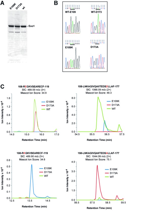
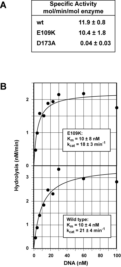
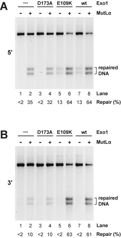
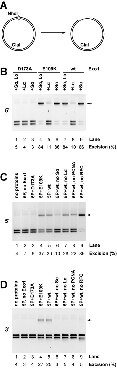
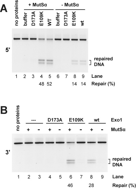
Genetic and biochemical studies have previously implicated exonuclease 1 (Exo1) in yeast and mammalian mismatch repair, with results suggesting that function of the protein in the reaction depends on both its hydrolytic activity and its ability to interact with other components of the repair system. However, recent analysis of an Exo1-E109K knockin mouse has concluded that Exo1 function in mammalian mismatch repair is restricted to a structural role, a conclusion based on a prior report that N-terminal His-tagged Exo1-E109K is hydrolytically defective. Because Glu-109 is distant from the nuclease hydrolytic center, we have compared the activity of untagged full-length Exo1-E109K with that of wild type Exo1 and the hydrolytically defective active site mutant Exo1-D173A. We show that the activity of Exo1-E109K is comparable to that of wild type enzyme in a conventional exonuclease assay and that in contrast to a D173A active site mutant, Exo1-E109K is fully functional in mismatch-provoked excision and repair. We conclude that the catalytic function of Exo1 is required for its participation in mismatch repair. We also consider the other phenotypes of the Exo1-E109K mouse in the context of Exo1 hydrolytic function.


Figures

Figure 1.
Figure 1.
Purity and sequence confirmation of Exo1 and Exo1 variants used in this study. (A) Samples (5 μg) of Exo1 preparations used in this study were subjected to polyacrylamide gel electrophoresis in the presence of sodium dodecyl sulfate and stained with Coomassie Brilliant Blue. (B) Lysates of baculovirus-infected SF9 cells employed for protein isolation were used as sources of template DNA for PCR amplification of the N-terminal 341 codons of Exo1. DNA sequence analysis of PCR products demonstrated presence of Glu and Asp codons at positions 109 and 173 for wild type Exo1 (upper reads); Lys at position 109 for Exo1-E109K (lower left read) and Ala at position 173 for Exo1-D173A (lower right read). (C) EIC peak traces of chymotryptic products spanning residues E109 and D173 across three LC-MS injections. Each EIC was performed ±20 ppm around the monoisotopic precursor m/z. Peaks were manually verified using retention time relative to qualitative peptide identification time as well as predicted ratio of C12 and C13 isotopomer peaks.
Figure 2.
Figure 2.
Exo1-E109K is a functional exonuclease. (A) Activities of wild type, Exo1-E109K and Exo1-D173A were determined as a function of enzyme concentration (0.01–2 nM) using a synthetic 5′-recessed 32P-labeled oligonucleotide duplex (27.5 nM, Materials and Methods). Specific activities shown were determined from progress curves where rates were linear with enzyme concentration. Results shown are the mean of 3 (wild type and Exo1-D173A) or 4 (Exo1-E109K) determinations (±one standard deviation). (B) Steady-state rates of [32P] synthetic duplex hydrolysis by 0.15 nM wild type Exo1 or Exo1-E109K were determined as a function of substrate concentration and results fit to a hyperbola using the non-linear regression function of DeltaGraph (RedRock Software). Km and kcat values shown in the insets are averages of three independent determinations (±one standard deviation).
Figure 3.
Figure 3.
The exonuclease function is required for Exo1-dependent mismatch repair in mouse cell extract. Mismatch repair was scored using G-T heteroduplex DNA containing a 5′- (panel A) or 3′- (panel B) strand break in whole cell extract prepared from Mlh1−/−Exo1−/− MEF cells. Extracts were supplemented with MutLα as indicated and assays performed in the absence (dilution buffer only) or presence of Exo1-D173A, Exo1-E109K or wild type Exo1 (Materials and Methods). Mismatch repair, which converts the heteroduplex to a HindIII-sensitive form, was determined after cleavage with HindIII and ClaI.
Figure 4.
Figure 4.
Mismatch-provoked excision in reconstituted systems requires the Exo1 hydrolytic function. (A) G-T heteroduplexes used in this study contain a NheI site that is located 5 bp distal to the mismatch in the 5′-heteroduplex and 5 bp proximal to the mispair in the 3′-heteroduplex. Mismatch provoked excision, which renders this region single-stranded and NheI-resistant (9,10), was scored by cleavage with NheI and ClaI. Arrows designate excision products in panels B–D. (B) 5′-directed excision on the G-T heteroduplex was determined in the 4-protein system. Reactions contained RPA with MutSα and MutLα present as indicated. Excision was scored in the presence of Exo1-D173A, Exo1-E109K or wild type enzyme (Materials and Methods). (C) 5′-directed excision in the 6-protein system was determined in reactions containing MutSα, MutLα, RPA, RFC, PCNA (indicated as 5P in lane labels), in the absence (lane 2) or presence of Exo1-D173A, Exo1-E109K or wild type Exo1 (lanes 3–5). Lanes 6–9 correspond to reactions containing wild type Exo1 with omissions as indicated. (D) Reactions were as in panel C except the heteroduplex strand break was located 3′ to the mismatch.
Figure 5.
Figure 5.
Exo1 hydrolytic action is required for reconstituted mismatch repair. Mismatch repair of 5′- (panel A) or 3′- (panel B) G-T heteroduplex DNAs was determined in the presence of MutLα, RPA, RFC, PCNA and DNA polymerase δ in the absence or presence of MutSα, Exo1-D173A, Exo1-E109K or wild type Exo1 as indicated. Mismatch repair was determined as in Figure 3.

References

    1. Tran P.T., Erdeniz N., Symington L.S., Liskay R.M. EXO1-A multi-tasking eukaryotic nuclease. DNA Repair (Amst) 2004;3:1549–1559.. - PubMed
    1. Szankasi P., Smith G.R. A DNA exonuclease induced during meiosis of Schizosaccharomyces pombe. J. Biol. Chem. 1992;267:3014–3023.. - PubMed
    1. Szankasi P., Smith G.R. A role for exonuclease I from S. pombe in mutation avoidance and mismatch correction. Science. 1995;267:1166–1169.. - PubMed
    1. Rudolph C., Fleck O., Kohli J. Schizosaccharomyces pombe exo1 is involved in the same mismatch repair pathway as msh2 and pms1. Curr. Genet. 1998;34:343–350.. - PubMed
    1. Dherin C., Gueneau E., Francin M., Nunez M., Miron S., Liberti S.E., Rasmussen L.J., Zinn-Justin S., Gilquin B., Charbonnier J.B., et al. Characterization of a highly conserved binding site of Mlh1 required for exonuclease I-dependent mismatch repair. Mol. Cell. Biol. 2009;29:907–918.. - PMC - PubMed

Publication types

Substances