. 2014 Jul 15;307(2):F183-94.
doi: 10.1152/ajprenal.00177.2014. Epub 2014 May 14.

Semaphorin 3A inactivation suppresses ischemia-reperfusion-induced inflammation and acute kidney injury

Affiliations

Semaphorin 3A inactivation suppresses ischemia-reperfusion-induced inflammation and acute kidney injury

Punithavathi Ranganathan et al. Am J Physiol Renal Physiol. .

Abstract

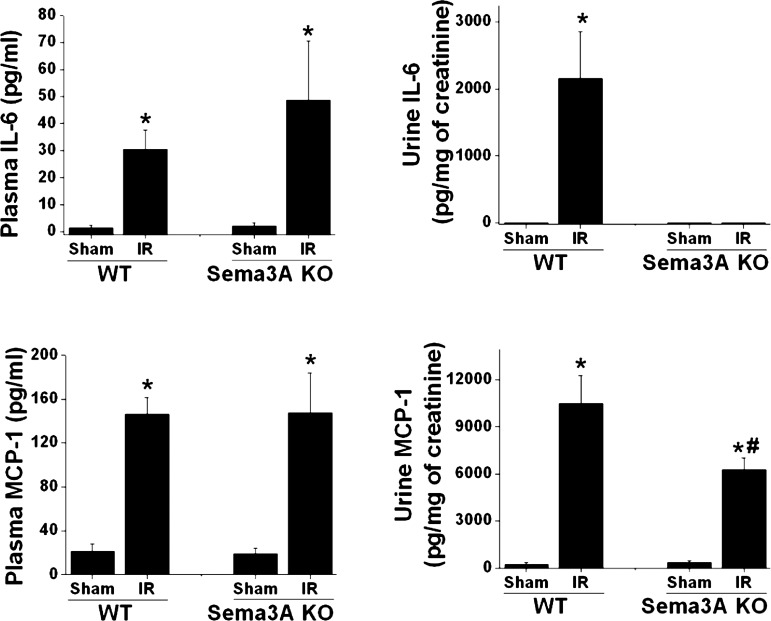
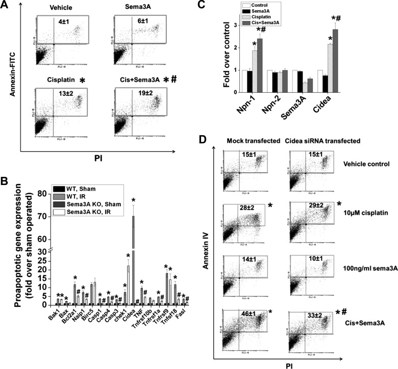
Recent studies show that guidance molecules that are known to regulate cell migration during development may also play an important role in adult pathophysiologic states. One such molecule, semaphorin3A (sema3A), is highly expressed after acute kidney injury (AKI) in mice and humans, but its pathophysiological role is unknown. Genetic inactivation of sema3A protected mice from ischemia-reperfusion-induced AKI, improved tissue histology, reduced neutrophil infiltration, prevented epithelial cell apoptosis, and increased cytokine and chemokine excretion in urine. Pharmacological-based inhibition of sema3A receptor binding likewise protected against ischemia-reperfusion-induced AKI. In vitro, sema3A enhanced toll-like receptor 4-mediated inflammation in epithelial cells, macrophages, and dendritic cells. Moreover, administration of sema3A-treated, bone marrow-derived dendritic cells exacerbated kidney injury. Finally, sema3A augmented cisplatin-induced apoptosis in kidney epithelial cells in vitro via expression of DFFA-like effector a (cidea). Our data suggest that the guidance molecule sema3A exacerbates AKI via promoting inflammation and epithelial cell apoptosis.

Keywords: acute kidney injury; cisplatin; semaphorin3A.


Figures

Fig. 1.
Fig. 1.
Regulation of expression of semaphorin3A (sema3A) and its receptors in response to ischemia-reperfusion (IR) of the kidney. A: immunohistochemical localization of sema3A and neuropilin-1 in mouse kidney. Sema3A (green) is localized in podocytes, and in distal and collecting duct epithelial cells, in sham-operated kidney, and the intensity of staining was increased following IR. Staining for sema3A was also detected in the injured proximal tubular epithelial cells after reperfusion injury. White asterisk shows megalin (red) and sema3A colocalization (bottom). Scale bar: 100 μM. B: sema3A receptor mRNA expression in wild-type (WT) and sema3A mutant mice in response to renal IR injury. Plexin-A1 was significantly upregulated, whereas plexin-A3 was downregulated in WT mice that were subjected to IR. In contrast, plexin-A2 expression was not altered. Neuropilin-1 expression was increased in WT mice following IR, which was strongly abrogated in sema3A mutant mice, while neuropilin-2 expression was unaffected by IR. *P < 0.05 vs. other groups; n = 4–6. KO, knockout.
Fig. 2.
Fig. 2.
Sema3A mutant mice were protected against IR-induced kidney injury. A: creatinine levels (index of renal function). Sema3A mutant or WT mice were subjected to 35 min of ischemia followed by 24 h of reperfusion. Sema3A mutant mice showed significantly lower serum creatinine compared with WT mice after 24 h of reperfusion. *P < 0.001 vs. sham-operated. #P < 0.05 vs. WT IR; n = 8–10. B: tissue histology for acute tubular necrosis, assessed in periodic acid-Schiff (PAS)-stained kidney sections. Sham-operated WT and sema3A mutant mice exhibited normal tubular structure. Following IR, kidneys from WT mice exhibited extensive tubular necrosis, loss of brush border, and cast formation. These changes were strongly abrogated in sema3A mutant mice. Quantification of tubular injury is shown in C. *P < 0.001 vs. sham-operated. #P < 0.001 vs. WT IR; n = 6–8.
Fig. 3.
Fig. 3.
IR-induced inflammatory mediators and glial cell line-derived neurotrophic factor (GDNF) expression are suppressed in sema3A mutant mice. IR induced a large increase in mRNA expression of proinflammatory mediators in the kidneys of WT mice, which was significantly suppressed in sema3A mutant mice. *P < 0.001 vs. other groups. #P < 0.001 vs. WT IR; n = 4–6.
Fig. 4.
Fig. 4.
Sema3A mutant mice exhibited reduced urinary excretion of IL-6 and MCP-1 following IR, while plasma levels were unchanged. IL-6 and MCP-1 in urine were quantified by ELISA as described in materials and methods and expressed as pg/mg of urine creatinine. *P < 0.001 vs. other groups. #P < 0.001 vs. WT IR; n = 5.
Fig. 5.
Fig. 5.
A: neutrophil infiltration into kidneys of WT and sema3A mutant mice subjected to sham operation or IR. Neutrophil infiltration was assessed by immunohistochemistry (top), and quantitative data are shown in bottom. *P < 0.001 vs. sham-operated. #P < 0.001 vs. WT IR; n = 4–6. B: apoptosis of tubular epithelial cells in kidneys of WT and sema3A mutant mice subjected to sham operation or IR. Apoptosis was assessed by TdT-mediated dUTP nick end labeling (TUNEL) staining. Blue nuclei indicate TUNEL-positive apoptotic cells (top). Quantitative data are shown in bottom. *P < 0.001 vs. sham-operated. #P < 0.001 vs. WT IR; n = 4–6.
Fig. 6.
Fig. 6.
LOXblock-1, an inhibitor of sema3A signaling, blocked kidney injury and prevented apoptosis in response to IR. A: kidney function was determined by measuring serum creatinine. *P < 0.001 vs. sham-operated. #P < 0.05 vs. IR vehicle treated. B: apoptosis of renal tubular epithelial cells was assessed by TUNEL staining. C: quantitative data for TUNEL-positive nuclei. *P < 0.001 vs. sham-operated. #P < 0.001 vs. IR vehicle treated; n = 4–6.
Fig. 7.
Fig. 7.
Sema3A-blocking peptide prevented kidney injury and blunted apoptosis in response to IR. A: kidney function was determined by measuring serum creatinine. *P < 0.001 vs. sham-operated. #P < 0.05 vs. IR scrambled peptide treated. B: apoptosis of renal tubular epithelial cells was assessed by TUNEL staining. C: quantitative data for TUNEL-positive nuclei. *P < 0.001 vs. sham-operated. #P < 0.001 vs. IR scrambled peptide treated; n = 6.
Fig. 8.
Fig. 8.
Sema3A exacerbates cisplatin-induced apoptosis in renal tubular epithelial cells. A: quantification of apoptosis (PI and annexin V staining followed by flow cytometry analysis) in mouse proximal tubular epithelial cells (TKPTS) treated with vehicle or 10 μM cisplatin with/without 100 ng/ml sema3A for 24 h. *P < 0.05 vs. vehicle. #P < 0.05 vs. cisplatin. B: expression of proapoptotic genes in kidneys of WT and sema3A mice following IR, determined by PCR microarray analyses. *P < 0.05 vs. sham-operated. #P < 0.05 vs. WT IR. C: effects of cisplatin with/without sema3A on neuropilin, sema3A, and cidea (cell death-inducing DNA fragmentation factor, α subunit-like effector A) expression in TKPTS cells determined by RT-PCR analysis. *P < 0.05 vs. control and sema3A. #P < 0.05 vs. cisplatin; n = 4–6. D: cidea mediates sema3A-induced augmentation of apoptosis in renal epithelial cells (TKPTS). Cells transfected with siRNA against cidea or mock-transfected TKPTS cells were treated with cisplatin + vehicle or cisplatin + sema3A (100 ng/ml) for 24 h. Apoptosis was determined as described above. *P < 0.05 vs. vehicle control. #P < 0.05 vs. mock-transfected cisplatin-treated; n = 6.
Fig. 9.
Fig. 9.
Sema3A synergizes with Toll-like receptor 4 (TLR4) signaling to enhance cellular inflammatory responses and kidney injury. TKPTS (A), macrophages (B), and dendritic cells (C–F) were treated with a low dose of LPS (1 ng/ml) with/without sema3A (100 ng/ml) for 24 h. Control cells were treated with vehicle (0.1% BSA in PBS). Cytokine expression was quantified by RT-PCR and ELISA. Induction of the proinflammatory cytokine IL-6 by LPS was augmented by sema3A treated in TKPTS cells (A), macrophages (B), and dendritic cells (C). *P < 0.05 vs. vehicle-treated. #P < 0.05 vs. LPS- or sema3A-treated. $P < 0.05 vs. LPS-treated. C: flow cytometry analysis of in vitro differentiated bone marrow cells. Ninety-eight percent of cells were positive for CD11c and CD11b, and negative for F4/80 monocyte/macrophage marker, consistent with myeloid-derived dendritic cells (DC). D: upregulation of cytokine expression in LPS-treated DC by sema3A. *P < 0.05 vs. vehicle-treated. #P < 0.05 vs. LPS-treated DC. E: sema3A increased expression of the DC activation marker CD40. *P < 0.001 vs. all other groups. F: administration of sema3A-treated DC before IR exacerbates kidney injury. *P < 0.05 vs. sham. #P < 0.05 vs. vehicle-treated DC; n = 6.
Fig. 10.
Fig. 10.
Infusion of sema3A-treated bone marrow-derived DC (BM-DC) before IR exacerbated kidney injury. Kidney sections were stained with PAS-hematoxylin, and tubular injury was scored as described in materials and methods. A: sham-operated kidney. B: infusion of vehicle-treated BM-DC before IR, showing tubular necrosis and cast formation. C: infusion of sema3A-treated BM-DC before IR, showing more extensive tubular necrosis and cast formation. D: quantification of tissue injury. *P < 0.01 vs. sham-operated. #P < 0.05 vs. vehicle-treated DC; n = 4. E: inflammatory cytokine and chemokine expression (analyzed by real-time RT-PCR) in kidneys of the 3 groups of mice. *P < 0.001 vs. sham. #P < 0.05 vs. vehicle-treated DC; n = 4.

Similar articles

Cited by

References

    1. Bagri A, Marin O, Plump AS, Mak J, Pleasure SJ. Slit proteins prevent midline crossing and determine the dorsoventral position of major axonal pathways in the mammalian forebrain. Neuron 33: 233–248, 2002 - PubMed
    1. Behar O, Golden JA, Mashimo H, Schoen FJ, Fishman MC. Semaphorin III is needed for normal patterning and growth of nerves, bones and heart. Nature 383: 525–528, 1996 - PubMed
    1. Bellomo R, Ronco C, Kellum JA, Mehta RL, Palevsky P. Acute renal failure–definition, outcome measures, animal models, fluid therapy and information technology needs: the Second International Consensus Conference of the Acute Dialysis Quality Initiative (ADQI) Group. Crit Care 8: R204–R212, 2004 - PMC - PubMed
    1. Bielenberg DR, Klagsbrun M. Targeting endothelial and tumor cells with semaphorins. Cancer Metastasis Rev 26: 421–431, 2007 - PubMed
    1. Bonventre JV, Zuk A. Ischemic acute renal failure: an inflammatory disease? Kidney Int 66: 480–485, 2004 - PubMed

Publication types

MeSH terms