. 2014 Jul 15;307(2):F205-21.
doi: 10.1152/ajprenal.00638.2013. Epub 2014 May 14.

Accelerated receptor shedding inhibits kidney injury molecule-1 (KIM-1)-mediated efferocytosis

Affiliations

Accelerated receptor shedding inhibits kidney injury molecule-1 (KIM-1)-mediated efferocytosis

Rushi Gandhi et al. Am J Physiol Renal Physiol. .

Abstract

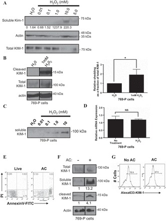
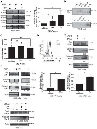
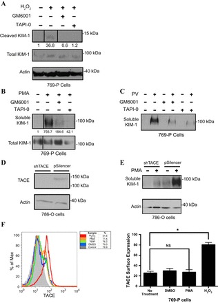
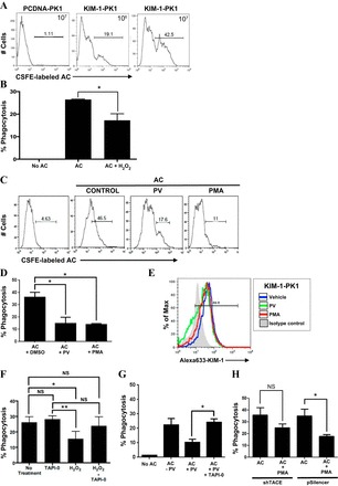
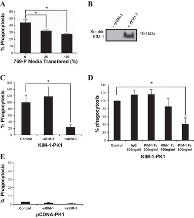
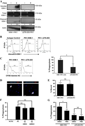
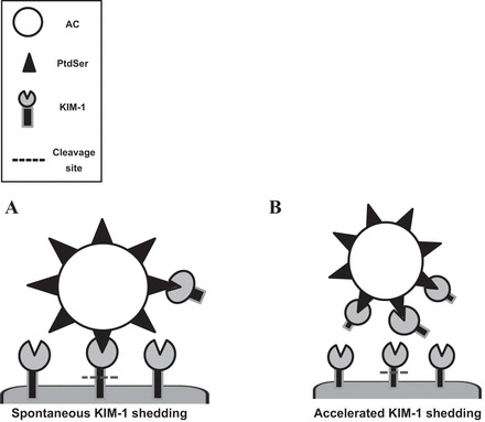
Efficient clearance of apoptotic cells (efferocytosis) prevents inflammation and permits repair following tissue injury. Kidney injury molecule-1 (KIM-1) is a receptor for phosphatidylserine, an "eat-me" signal exposed on the surface of apoptotic cells that marks them for phagocytic clearance. KIM-1 is upregulated on proximal tubule epithelial cells (PTECs) during ischemic acute kidney injury (AKI), enabling efferocytosis by surviving PTECs. KIM-1 is spontaneously cleaved at its ectodomain region to generate a soluble fragment that serves a sensitive and specific biomarker for AKI, but the biological relevance of KIM-1 shedding is unknown. Here, we sought to determine how KIM-1 shedding might regulate efferocytosis. Using cells that endogenously and exogenously express KIM-1, we found that hydrogen peroxide-mediated oxidative injury or PMA treatment accelerated KIM-1 shedding in a dose-dependent manner. KIM-1 shedding was also accelerated when apoptotic cells were added. Accelerated shedding or the presence of excess soluble KIM-1 in the extracellular milieu significantly inhibited efferocytosis. We also identified that TNF-α-converting enzyme (TACE or ADAM17) mediates both the spontaneous and PMA-accelerated shedding of KIM-1. While accelerated shedding inhibited efferocytosis, we found that spontaneous KIM-1 cleavage does not affect the phagocytic efficiency of PTECs. Our results suggest that KIM-1 shedding is accelerated by worsening cellular injury, and excess soluble KIM-1 competitively inhibits efferocytosis. These findings may be important in AKI when there is severe cellular injury.

Keywords: efferocytosis; kidney injury molecule-1 (KIM-1); proximal tubule epithelial cells (PTECs); receptor; shedding.


Figures

Fig. 1.
Fig. 1.
H2O2 and excess apoptotic cells accelerate Kidney injury molecule-1 (KIM-1) shedding. A: primary mouse proximal tubule epithelial cells (PTECs) were treated with increasing concentrations of H2O2, and both conditioned media and total cell lysates were collected. Soluble mouse KIM-1 was detected by SDS-PAGE and Western blotting. Soluble KIM-1 relative to total KIM-1 was quantified by densitometry. B: 769-P cells were exposed to H2O (1 μl) or H2O2 (1 mM) for 30 min in serum-free DMEM and the total cell lysates were collected and analyzed by Western blotting for cleaved and total KIM-1. The graph summarizes cleaved KIM-1 relative to total KIM-1 as determined by densitometry from 3 independent experiments (right). C: confluent monolayers of 769-P cells plated in 6-well tissue culture plates were exposed to H2O (1 μl) or various concentrations of H2O2 (0.01, 0.1, 1.0, and 10 mM) for 30 min in serum-free DMEM before analysis of the conditioned medium for soluble KIM-1. D: H2O2-induced KIM-1 shedding occurs independently of changes in KIM-1 mRNA expression. 769-P cells were exposed to H2O (1 μl) or H2O2 (1 mM) for 30 min in serum-free DMEM. Total cellular RNA was harvested, and relative KIM-1 mRNA expression was quantified using quantitative RT-PCR. E: UV-induced apoptotic(AC) or live mouse thymocytes that were stained with annexin V-FITC and propidium iodide (PI) and analyzed by flow cytometry. F: confluent monolayers of 769-P cells were not exposed or exposed to 107 apoptotic cells for 1 h. Relative shedding as a fraction of total KIM-1 was quantified by densitometry. A–E: soluble KIM-1 was detected from media samples with AKG antibody. Cleaved and total KIM-1 were detected in the total cellular lysates with 195 antibody. Actin was used as a loading control. The Western blot result is representative of 3 independent experiments. G: after being not fed or fed 5-(and 6)-carboxyfluorescein di-acetate succinimidyl ester (CFSE)-labeled apoptotic cells (AC) as in D, single-cell suspensions of the 769-P cells were analyzed for change in surface KIM-1 expression (Alexa 633-KIM-1) after staining with anti-KIM-1 primary antibody (AKG) and Alexa 633-conjugated secondary antibody. NS, not significant. *P < 0.05.
Fig. 2.
Fig. 2.
PMA and pervanadate (PV) accelerate KIM-1 shedding. A: confluent monolayers of 769-P cells were incubated in serum-free DMEM with DMSO (control) or PMA, (1 μM) for 1 h or PV (50 μM) for 30 min. A graph summarizing soluble KIM-1 relative to total KIM-1 in 769-P cells as determined by densitometry from 3 independent experiments is shown. B: 769-P cells were pretreated with PMA at various concentrations (0, 10, and 100 nM, and 5 μM) for 30 min (left) and for various durations of time (30 min, 1 h, or 2 h) at a concentration of 1 μM (right). C: PMA-induced KIM-1 shedding occurs independently of mRNA synthesis. 769-P cells were exposed to DMSO (control) or PMA for 1 h in serum-free DMEM. Total cellular RNA was harvested, and relative KIM-1 mRNA expression was analyzed using quantitative RT-PCR. D: 769-P cells were incubated for 30 min with or without PMA (1 μM) or PV (50 μM) before detection of surface KIM-1 expression by flow cytometry using AKG antibody and Alexa Fluoro 633-conjugated as secondary antibodies. KIM-1 expression is displayed in the form of a single-parameter (Alex 633-KIM-1) histogram against the % maximum mean fluorescent intensity. E: PMA-induced KIM-1 shedding occurs independently of protein synthesis. 769-P cells were preincubated for 30 min with cycloheximide (CHX; 25 μg/ml) in DMEM medium and then, with DMSO (control) or PMA (1 μM) for an additional 1 h before detection of soluble, cleaved, and total KIM-1 by Western blotting. F: KIM-1-PK1 cells were incubated in DMEM with DMSO (control) or PMA (1 μM) for 1 h, or with or without PV (50 μM) for 30 min. Graph summarizing cleaved KIM-1 relative total KIM-1 in KIM-1-PK-1 cells as determined by densitometry from 3 independent experiments is shown. G: HK-2 cells were incubated in DMEM with DMSO (control) or PMA (1 μM) for 1 h, or PV (50 μM) for 30 min. A–G: soluble KIM-1 was detected from media samples with AKG antibody. Cleaved and total KIM-1 were detected in the total cellular lysates with 195 antibody. Actin was used as a loading control. Cleaved KIM-1 relative to total KIM-1 was quantified by densitometry. Western blot results described above are representative of results obtained from 3 independent experiments. *P < 0.05.
Fig. 3.
Fig. 3.
Constitutive and accelerated KIM-1 shedding is inhibited by TNF-α-converting enzyme (TACE) inhibitors and short hairpin (sh) RNA-mediated knockdown of TACE. A: confluent monolayers of 769-P renal cell carcinoma (RCC) cells were first pretreated with DMSO (control), GM6001 (1 μM), or TAPI-0 (10 μM) for 30 min following exposure to H2O (1 μl) or H2O2 (1 mM) for 30 min. Shed KIM-1 was detected in media with AKG antibody. Cleaved and total KIM-1 was detected in total cellular lysate with 195 antibody. B and C: experiment in A was repeated with PMA (1 μM) for 1 h and PV (50 μM) for 30 min. A and B: soluble KIM-1 relative to total KIM-1 was quantified by densitometry. Lines between the bands represent any lanes that were rearranged from the original blot. Cleaved KIM-1 was detected using a 15% gel, while total KIM-1 and actin were detected using a 10% gel. D: cell lysates of 786-O RCC cells stably expressing a vector encoding shRNA targeting TACE (shTACE) or the empty vector alone (pSilencer) were analyzed by Western blotting with anti-TACE antibody. A band corresponding to TACE is shown. E: confluent monolayers shTACE and pSilencer cells were preincubated with PMA for 30 min, and KIM-1 shedding was assessed by Western blot analysis. Actin was used as a loading control. The Western blot data are representative of the results obtained from 3 independent experiments. F: 769-P cells were incubated with H2O (1 μl), H2O2 (1 mM), DMSO, or PMA (1 μM) for 30 and 60 min, respectively, before detection of surface TACE expression by flow cytometry using a TACE antibody. Surface TACE expression is displayed in the form of a histogram against the % maximum mean fluorescent intensity and as a graph representative of data from 3 independent experiments. *P < 0.05.
Fig. 4.
Fig. 4.
Accelerated KIM-1 shedding inhibits efferocytosis in KIM-1-expressing PTECs. A: PCDNA-PK1 or KIM-1-PK1 cells were incubated with 106 or 107 CFSE-labeled apoptotic cells(AC) in 6-well plates for 90 min, and the phagocytic uptake of apoptotic cells was analyzed by flow cytometry. Data re depicted in the form of a representative single-parameter (CFSE-labeled AC) histogram determined from 3 independent experiments. B: KIM-1-PK1 cells were untreated or pretreated with H2O (control) or H2O2 (1 mM) for 30 min in 6-well plates followed by incubation with 107 CFSE-labeled apoptotic cells for 90 min before measurement of phagocytic uptake of apoptotic cells by flow cytometry. C: KIM-1-PK1 cells were pretreated with DMSO (control), PV (50 μM) for 30 min, or PMA (1 μM) for 1 h in 6-well plates followed by incubation with 107 CFSE-labeled apoptotic cells for 90 min. Samples were analyzed by flow cytometry as in A. D: data from 3 independent experiments in C were graphed to show percent phagocytosis with error bars representing SD. E: change in surface KIM-1- expression in KIM-1-PK1 cells after addition of DMSO (vehicle), PMA, and PV as determined by flow cytometry using AKG primary antibody and Alexa Fluoro 633-conjugated anti-mouse IgG secondary antibody. F: 769-P cells were first pretreated with TAPI-0 (10 μM) for 30 min and then with H2O2 (1 mM) for 30 min before incubation with 106 CFSE-labeled apoptotic cells for an additional 90 min. Percent phagocytosis was determined as described in A from 3 independent experiments. G: KIM-1-PK1 cells were first pretreated with TAPI-0 (10 μM) for 30 min and then with DMSO (−PV) or PV (+PV) for another 30 min before incubation with 106 CFSE-labeled apoptotic cells for an additional 90 min. Percent phagocytosis was determined as described in A. H: shTACE and pSilencer cells were pretreated with and without PMA (1 μM) for 1 h followed by incubation with apoptotic cells for 90 min. Flow cytometry analysis was done as in A. *P < 0.05.
Fig. 5.
Fig. 5.
Soluble KIM-1 competitively inhibits apoptotic cell engulfment by KIM-1-expressing PTECs. A: CFSE-labeled apoptotic cells were incubated on ice for 30 min with increasing fractions of conditioned medium from 769-P cells cultured for 3 days. Thereafter, the conditioned medium containing the apoptotic cells was added to confluent monolayers of KIM-1-PK1 cells for 90 min before measurement of phagocytic uptake by flow cytometry. B: Western blot detecting soluble KIM-1 in the conditioned medium collected from KIM1-Tet-off MDCKII cells either treated with doxycycline (100 ng/ml) to inhibit KIM-1 expression (−sKIM-1) or no doxycycline for 5 days, permitting high-level expression of KIM-1 (+sKIM-1). C: CFSE-labeled apoptotic cells were incubated on ice for 30 min with conditioned medium from KIM1-Tet-off MDCKII cells either treated with doxycycline (−sKIM-1) or no doxycycline (+sKIM-1). Thereafter, the conditioned media containing the apoptotic cells were added to monolayers of KIM-1-PK1 cells for 90 min before measurement of phagocytic uptake by flow cytometry. D: relative phagocytic uptake of labeled apoptotic cells by KIM-1-PK1 cells incubated in the absence or presence of recombinant KIM-1-Fc protein (n = 3) or nonspecific IgG at the concentration indicated. C and D: results are expressed as percent relative phagocytosis of untreated control. A–D: fresh culture medium was used as the control. E: soluble KIM-1 does not confer on PTECs the ability to engulf apoptotic cells in the absence of KIM-1 expression. CFSE-labeled apoptotic cells were incubated on ice for 30 min with the conditioned (−sKIM-1 or +sKIM-1) medium from KIM1-Tet-off MDCKII cells, and thereafter the conditioned media containing the apoptotic cells were added to the PCDNA-PK1 cells for an additional 90 min before measurement of phagocytic uptake by flow cytometry. *P < 0.05.
Fig. 6.
Fig. 6.
Phagocytic uptake of apoptotic cells is impaired in PTECs expressing a cleavage-mutant of KIM-1. A: LLC-PK1 cells stably expressing wild-type (KIM-1-PK1) or a cleavage-mutant of KIM-1 (Δ278–283-PK1) were treated with PMA (1 μM) or vehicle control (DMSO) for 30 min. Soluble and cleaved KIM-1 were detected from media and cell lysate samples, respectively. Actin was used as a loading control. Soluble and cleaved KIM-1 relative to total KIM-1 was quantified by densitometry. B: surface expression of KIM-1 in KIM-1-PK1 and Δ278–283-PK1 cells was evaluated by flow cytometry using both AKG or control antibody and Alexa Fluoro 633-conjugated secondary without permeabilization. C: KIM-1-PK1 and Δ278–283-PK1 cells were incubated with 106 CFSE-labeled apoptotic cells for 90 min, and phagocytosis was analyzed by flow cytometry. Data are depicted in the form of a representative single-parameter histogram (left) and a graph summarizing the relative (%) phagocytosis determined from 3 independent experiments (right). D: binding of pHrodo-labeled apoptotic cells(AC) to confluent monolayers of KIM-1-PK1 and Δ278–283-PK1 cells at 4°C as observed by confocal microscopy. Images were captured at 400× magnification, and the scale bar represents 10 μm. E: percent bound apoptotic cells relative to the total number of nuclei determined from 3 independent experiments in D. F: KIM-1-PK1 cells were pretreated with DMSO (control) or GM6001 (1 μM) for 30 min followed by incubation with 107 CFSE-labeled apoptotic cells (AC) for 90 min. Flow cytometric analysis was used to determine the percent phagocytosis from 3 independent experiments. G: KIM-1-PK1 and Δ278–283-PK1 cells were pretreated with DMSO (control) or PMA (1 μM) for 1 h in 6-well plates followed by incubation with 107 CFSE-labeled apoptotic cells for 90 min. Percent phagocytosis was determined by flow cytometry from 3 independent experiments. *P < 0.05.
Fig. 7.
Fig. 7.
Hypothetical model showing the regulation of KIM-1-mediated efferocytosis by receptor shedding. A: spontaneous KIM-1 shedding does not affect the efficiency of KIM-1-dependent efferocytosis. B: competitive inhibition of efferocytosis by decoy (soluble KIM-1) receptors resulting from excess KIM-1 shedding due to exaggerated cellular injury (e.g., H2O2).

Similar articles

Cited by

References

    1. Anderson HA, Maylock CA, Williams JA, Paweletz CP, Shu H, Shacter E. Serum-derived protein S binds to phosphatidylserine and stimulates the phagocytosis of apoptotic cells. Nat Immunol 4: 87–91, 2003. - PubMed
    1. Arribas J, Coodly L, Vollmer P, Kishimoto TK, Rose-John S, Massague J. Diverse cell surface protein ectodomains are shed by a system sensitive to metalloprotease inhibitors. J Biol Chem 271: 11376–11382, 1996. - PubMed
    1. Arribas J, Esselens C. ADAM17 as a therapeutic target in multiple diseases. Curr Pharm Des 15: 2319–2335, 2009. - PubMed
    1. Arroyo A, Modriansky M, Serinkan FB, Bello RI, Matsura T, Jiang J, Tyurin VA, Tyurina YY, Fadeel B, Kagan VE. NADPH oxidase-dependent oxidation and externalization of phosphatidylserine during apoptosis in Me2SO-differentiated HL-60 cells. Role in phagocytic clearance. J Biol Chem 277: 49965–49975, 2002. - PubMed
    1. Atabai K, Jame S, Azhar N, Kuo A, Lam M, McKleroy W, Dehart G, Rahman S, Xia DD, Melton AC, Wolters P, Emson CL, Turner SM, Werb Z, Sheppard D. Mfge8 diminishes the severity of tissue fibrosis in mice by binding and targeting collagen for uptake by macrophages. J Clin Invest 119: 3713–3722, 2009. - PMC - PubMed

Publication types

MeSH terms