. 2013 Aug 27:1:14.
doi: 10.1186/2051-1426-1-14. eCollection 2013.

PET imaging to non-invasively study immune activation leading to antitumor responses with a 4-1BB agonistic antibody

Affiliations

PET imaging to non-invasively study immune activation leading to antitumor responses with a 4-1BB agonistic antibody

Helena Escuin-Ordinas et al. J Immunother Cancer. .

Abstract

Background: Molecular imaging with positron emission tomography (PET) may allow the non-invasive study of the pharmacodynamic effects of agonistic monoclonal antibodies (mAb) to 4-1BB (CD137). 4-1BB is a member of the tumor necrosis factor family expressed on activated T cells and other immune cells, and activating 4-1BB antibodies are being tested for the treatment of patients with advanced cancers.

Methods: We studied the antitumor activity of 4-1BB mAb therapy using [(18) F]-labeled fluoro-2-deoxy-2-D-glucose ([(18) F]FDG) microPET scanning in a mouse model of colon cancer. Results of microPET imaging were correlated with morphological changes in tumors, draining lymph nodes as well as cell subset uptake of the metabolic PET tracer in vitro.

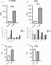
Results: The administration of 4-1BB mAb to Balb/c mice induced reproducible CT26 tumor regressions and improved survival; complete tumor shrinkage was achieved in the majority of mice. There was markedly increased [(18) F]FDG signal at the tumor site and draining lymph nodes. In a metabolic probe in vitro uptake assay, there was an 8-fold increase in uptake of [(3)H]DDG in leukocytes extracted from tumors and draining lymph nodes of mice treated with 4-1BB mAb compared to untreated mice, supporting the in vivo PET data.

Conclusion: Increased uptake of [(18) F]FDG by PET scans visualizes 4-1BB agonistic antibody-induced antitumor immune responses and can be used as a pharmacodynamic readout to guide the development of this class of antibodies in the clinic.

Keywords: 4-1BB; CD137; Colon cancer; Immune activating antibodies; PET imaging.


Figures
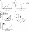
Figure 1
Figure 1
Antitumor activity of 4-1BB mAb therapy against CT26 colon carcinoma. a) The administration of a double dose of 4-1BB mAb (1 mg/kg) induced reproducible CT26 tumor regressions (n = 12 mice per group; 3.8 ± 0.4 4-1BB mAb group vs 9.2 ± 0.3 control, on day 14 post-tumor implant; p < 0.0001 by two-way Anova). b) Kaplan-Meier plot of improved survival in mice (n = 12 mice per group; p = 0.0001 for tumor diameters by two-way Anova and p = 0.0001 for survival by log rank test). c and d) Different 4-1BB mAb doses were also tested showing a significant tumor growth inhibition by day 21 in animals receiving 0.1 or 1 mg/kg 4-1BB mAb (ns = not significant * P < 0.05, ** P < 0.01, *** P < 0.001 by t-test). e) Re-challenge of surviving tumor-free mice 55 days after complete tumor shrinkage showed a total CT26 tumor rejection by the animals. 4 T1 cells were used as positive control.
Figure 2
Figure 2
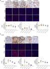
Pathological analysis of immune infiltration after 4-1BB mAb therapy. Tumor CD3+, F4/80+ and CD45+ infiltrating cells were identified by immunohistochemical and immunoflurecence analysis on day 14 (a) and day 22 (b) after tumor injection. Data is also represented by pixel area of positively stained cells per 100 pixel area of viable tumor. Error bars represent mean ± SEM, *p < 0.05.
Figure 3
Figure 3
Effects of 4-1BB agonistic antibody on the T cell functional phenotype. Peripheral blood mononuclear cells were stained with antibodies to CD3, CD4, CD8, CD44 and CD62L on day 14 or day 22 post-tumor implant. CD3 + CD8+ and CD3 + CD4+ T cells were further evaluated for T central memory (Tcm) (CD44 + CD62L+), T effector memory (Tem) (CD44 + CD62L-), and T naïve (CD44loCD62Lhi) cell subsets. The ratio of CD8 to CD4 T cells is shown (a). The percentage of CD4 and CD8 T cell populations in treated versus untreated mice is also shown (a, b, c) as well as the percentages of the different CD4 and CD8 cell subsets: Tcm (e, h), Tem (f, i) and T naïve cells (d, g), represented as a percentage of the parental population gate. *p < 0.05, **p < 0.01, ***p < 0.005 by t-test, n ≥ 3 mice/group, data is representative of 3 independent experiments.
Figure 4
Figure 4
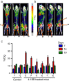
PET imaging of anti-4-1BB treatment in CT26 tumor-bearing mice. [18 F]FDG microPET imaging showed an increased signal (% ID/g) of [18 F]FDG at the tumor site and draining lymph nodes (DLNs) of mice treated with 4-1BB mAb (19.1 ± 2.1 4-1BB mAb group vs. 10.3 ± 0.11 control group, p = 0.06). Representative images shown; orange arrows indicate tumor (T) circled in white, and bladder (Bl). Control mice (a) and 4-1BB mAb treated mice (b) on day 6 (left), 14 (middle) and 19 (right) post-tumor implant. All mice were treated with 4-1BB mAb or saline on day 9 and 11 post-tumor inoculation. Muscle serves as a background control. (c) Standardized uptake values (SUVs) of all the imaged mice. Mouse B is slightly rotated in order for the tumor FDG uptake signal not to overlap with the bladder uptake signal.
Figure 5
Figure 5
In vitro uptake assay of [3H]DDG in cells obtained from mice treated with anti-4-1BB. Cells were extracted on day 14 post-tumor implant from mice bearing CT26 tumors treated with 4-1BB mAb, which showed an 8-fold increase in non-adherent CD45+ cells extracted from tumors (TI_CD45+; a) and draining lymph nodes (DLN; b) of mice treated with 4-1BB mAb compared to untreated mice. The TI_CD45+ and DLN cell populations are represented in bar graphs (c, d). e)In vitro co-culture of CT26 cells and splenocytes extracted from the CT26 tumor bearing mice, also showed a significant 2-fold increase in uptake of [3H]DDG in splenocytes isolated from the treated mice compared to the untreated mice. CT26 tumor cell uptake did not show a significant difference between the group treated with 4-1BB mAb and the untreated group (f). ** p < 0.01 and *** p < 0.0001 by t-test.

References

    1. Melero I, Shuford WW, Newby SA, Aruffo A, Ledbetter JA, Hellstrom KE, Mittler RS, Chen L. Monoclonal antibodies against the 4-1BB T-cell activation molecule eradicate established tumors. Nat Med. 1997;1(6):682–685. doi: 10.1038/nm0697-682. - DOI - PubMed
    1. Melero I, Hervas-Stubbs S, Glennie M, Pardoll DM, Chen L. Immunostimulatory monoclonal antibodies for cancer therapy. Nat Rev Cancer. 2007;1(2):95–106. doi: 10.1038/nrc2051. - DOI - PubMed
    1. Vinay DS, Kwon BS. Role of 4-1BB in immune responses. Semin Immunol. 1998;1(6):481–489. doi: 10.1006/smim.1998.0157. - DOI - PubMed
    1. Vinay DS, Kwon BS. Immunotherapy of cancer with 4-1BB. Mol Cancer Ther. 2012;1(5):1062–1070. doi: 10.1158/1535-7163.MCT-11-0677. - DOI - PubMed
    1. Wang C, Lin GH, McPherson AJ, Watts TH. Immune regulation by 4-1BB and 4-1BBL: complexities and challenges. Immunol Rev. 2009;1(1):192–215. doi: 10.1111/j.1600-065X.2009.00765.x. - DOI - PubMed