

Orbital Tumors Excision without Bony Marginotomy under Local and General Anesthesia
- PMID: 24829795
- PMCID: PMC4009292
- DOI: 10.1155/2014/424852
Orbital Tumors Excision without Bony Marginotomy under Local and General Anesthesia
Abstract
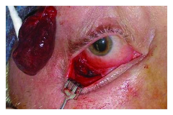
To present our experience of removing middle to deep orbital tumors using a combination of minimally invasive soft tissue approaches, sometimes under local anesthesia. Methods. In this retrospective case series, 30 patients (13 males and 17 females) underwent tumor removal through eyelid crease (17 eyes), conjunctival (nine eyes), lateral canthal (two eyes), and transcaruncular (two eyes) approaches. All tumors were located in the posterior half of the orbit. Six cases were removed under monitored anesthesia care with local block, and 24 were under general anesthesia. Results. The median (range) age and follow-up duration were 48.5 (31-87) years old and 24.5 (4-375) weeks, respectively. Visual acuity and ocular motility showed improvement or no significant change in all but one patient at the latest followup. Confirmed pathologies revealed cavernous hemangioma (15 cases), pleomorphic adenoma (5 cases), solitary fibrous tumor (4 cases), neurofibroma (2 cases), schwannoma (2 cases), and orbital varix (1 case). None of the patients experienced recurrence. Conclusions. Creating a bony marginotomy increases intraoperative exposure of the deep orbit but adds substantial time and morbidity. Benign orbital tumors can often be removed safely through small soft-tissue incisions, without bone removal and under local anesthesia.
Figures


References
-
- Burroughs JR, Soparkar CNS, Patrinely JR, Kersten RC, Kulwin DR, Lowe CL. Monitored anesthesia care for enucleations and eviscerations. Ophthalmology. 2003;110(2):311–313. - PubMed
-
- Kumar C. Orbital regional anesthesia: complications and their prevention. Indian Journal of Ophthalmology. 2006;54(2):77–84. - PubMed
-
- Leatherbarrow B, Noble JL, Lloyd IC. Cavernous haemangioma of the orbit. Eye. 1989;3(1):90–99. - PubMed
-
- Schick U, Dott U, Hassler W. Surgical treatment of orbital cavernomas. Surgical Neurology. 2003;60(3):234–244. - PubMed
-
- Thorn-Kany M, Arrué P, Delisle MB, Lacroix F, Lagarrigue J, Manelfe C. Cavernous hemangiomas of the orbit: MR imaging. Journal of Neuroradiology. 1999;26(2):79–86. - PubMed
LinkOut - more resources
Full Text Sources
Other Literature Sources
