. 2014:2014:670756.
doi: 10.1155/2014/670756. Epub 2014 Apr 16.

Plastic changes in the spinal cord in motor neuron disease

Affiliations

Plastic changes in the spinal cord in motor neuron disease

Francesco Fornai et al. Biomed Res Int. 2014.

Abstract

In the present paper, we analyze the cell number within lamina X at the end stage of disease in a G93A mouse model of ALS; the effects induced by lithium; the stem-cell like phenotype of lamina X cells during ALS; the differentiation of these cells towards either a glial or neuronal phenotype. In summary we found that G93A mouse model of ALS produces an increase in lamina X cells which is further augmented by lithium administration. In the absence of lithium these nestin positive stem-like cells preferentially differentiate into glia (GFAP positive), while in the presence of lithium these cells differentiate towards a neuron-like phenotype ( β III-tubulin, NeuN, and calbindin-D28K positive). These effects of lithium are observed concomitantly with attenuation in disease progression and are reminiscent of neurogenetic effects induced by lithium in the subependymal ventricular zone of the hippocampus.


Figures

Figure 1
Figure 1
Effects of lithium on survival and motor performance in G93A mice. The graphs report data from G93A mice with either saline or lithium. Lithium prolongs mice survival as shown by Kaplan-Meier survival curve. Lithium counteracts motor deterioration measured by stride length, paw grip endurance (PaGE), and rotarod. Values are given as the mean ± SEM. *P ≤ 0.05 compared with mice administered saline.
Figure 2
Figure 2
Pilot light microscopy procedures showing semithin sections. For identification of lamina X and ependymal area, 1-2 μm thick serial sections, obtained with a porter blum MT-1 or an ultramicrotome, were stained with 1% toluidine blue and 1% methylene blue in 1% sodium tetraborate and observed under light microscopy. In each experimental group (wild type administered saline, WT Sal; wild type administered lithium, WT Li; transgenic SOD1 G93A mice administered saline, G93A Sal; transgenic SOD1 G93A mice administered lithium, G93A Li) the central canal is limited by densely packed cells with polymorphic nuclei, which increase their number and appear as multiple call layers in G93A mice compared with WT. Again, in G93A mice, the staining is consistently more intense compared with WT. Scale bar = 21 μm.
Figure 3
Figure 3
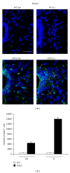
Nestin immunostaining within lamina X. Representative pictures are reported in (a) showing that lamina X shows neglectable nestin-immunofluorescence in WT mice (both WT Sal or WT Li). In contrast, nestin immunostaining is well evident within the lamina X from ALS mice (both G93A Sal and G93A Li). In particular, in G93A mice treated with saline nestin-positive fibers between the ependymal cells appear to project from the ependymal canal into lamina X, where only a few cells appear labelled. In contrast, in G93A mice treated with lithium nestin immunofluorescence is dramatic and involves both the ependymal cells and stem-like cells within the entire lamina X (arrows). (b) The graph reports the count of nestin-positive cells in both lamina X and ependymal canal. *P ≤ 0.05 compared with other groups. Scale bar = 26 μm.
Figure 4
Figure 4
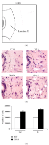
H&E staining of lamina X. (a) Atlas plate [25] of the lumbar cord (L4) used as the reference for representative pictures. (b) Representative high magnification of H&E stained pictures showing L4 lamina X, treated with either saline or lithium. The cell number counted within lamina X is equivalent for WT mice treated with either saline (WT Sal) or lithium (WT Li). On the other hand, the cell number is increased in the lamina X of G93A mice treated with saline (G93A Sal) or lithium (G93A Li). When counting the cell number it is evident that, as reported in the histogram (c), the cell number in lamina X is mostly increased in G93A mice receiving lithium. The increase in the cell number occurring within lamina X of G93A mice is better visualized at the level of the ependymal layer. Dashed lines draw the border of the lamina X. *P ≤ 0.05 compared with other groups. Scale bar = 30 μm.
Figure 5
Figure 5
Representative pictures from hemi-lumbar cord. Representative pictures of H&E stained hemi-lumbar cord. In the upper lane are reported pictures from wild type mice treated with either saline (WT Sal) or lithium (WT Li). In the lower lane are reported G93A mice treated with either saline (G93A Sal) or lithium (G93A Li). Pictures are representative of end stage of disease. Noteworthy is the generalized increase in spinal cord cell density which is produced by lithium administration in G93A mice. Such an effect appears to extend in the whole grey matter. Scale bar = 232 μm.
Figure 6
Figure 6
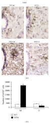
GFAP immunostaining within lamina X. Representative high magnification of GFAP immunostained slices from lamina X of lumbar spinal cord (a). The cell number counted within lamina X is equivalent for WT mice treated with either saline (WT Sal) or lithium (WT Li). On the other hand, the cell number is dramatically increased in the lamina X of G93A mice treated with saline (G93A Sal), while it is suppressed in G93A mice treated with lithium (G93A Li) as reported in the histogram (b). In general, GFAP immunostaining shows positive cells on external side of the ependymal layer, while in G93A Sal intense immunostaining extends in the surrounding lamina X. *P ≤ 0.05 compared with other groups. Scale bar = 30 μm.
Figure 7
Figure 7
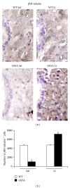
βIII-tubulin immunostaining within lamina X. Representative high magnification of βIII-tubulin immunostained slices from lamina X of lumbar spinal cord (a). The cell number counted within lamina X is equivalent for WT mice treated with either saline (WT Sal) or lithium (WT Li). On the other hand, the cell number is dramatically increased in the lamina X of G93A mice treated with lithium (G93A Li), while it is suppressed in G93A mice treated with saline (G93A Sal) as reported in the histogram (b). In general, βIII-tubulin immunostaining shows an opposite pattern compared with GFAP (see Figure 6). Following lithium administration an intense immunostaining is visible throughout the whole lamina X of G93A mice. *P ≤ 0.05 compared with other groups. Scale bar = 30 μm.
Figure 8
Figure 8
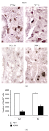
NeuN immunostaining within lamina X. Representative high magnification of NeuN immunostained slices from lamina X of lumbar spinal cord (a). Again, immunopositive cells within lamina X for WT mice treated with either saline or lithium (WT Sal or WT Li, resp.) is equivalent. On the other hand, NeuN positive cells are suppressed in the lamina X of saline treated G93A mice (G93A Sal), while lithium administration (G93A Li) partially recovers NeuN immunostaining as reported in the histogram (b). *P ≤ 0.05 compared with other groups. Scale bar = 30 μm.
Figure 9
Figure 9
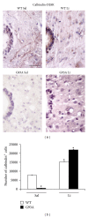
Calbindin-D28K immunostaining within lamina X. Representative high magnification of calbindin-D28K immunostaining within lamina X of lumbar spinal cord (a). In general, intense calbindin-D28K immunopositivity appears to be localized in the ependymal layer. The cell number counted within lamina X is markedly increased by lithium in both WT and G93A mice (WT Li and G93A Li, resp.). On the other hand, the cell number is reduced in G93A mice administered saline (G93A Sal). This latter effect is particularly evident in the area of lamina X external to the ependymal canal. In this area only pale cell shapes avoided from the count are visible, which explains the very low cell number reported in the histogram (b). In G93A mice the effects of lithium extend the strong immunostaining to the entire lamina X. *P ≤ 0.05 compared with other groups. Scale bar = 36 μm.

References

    1. Han Q, Feng J, Qu Y, et al. Spinal cord maturation and locomotion in mice with an isolated cortex. Neuroscience. 2013;253:235–244. - PubMed
    1. Johnson RA, Mitchell GS. Common mechanisms of compensatory respiratory plasticity in spinal neurological disorders. Respiratory Physiology and Neurobiology. 2013;189(2):419–428. - PMC - PubMed
    1. Bradley WG. Recent views on amyotrophic lateral sclerosis with emphasis on electrophysiological studies. Muscle and Nerve. 1987;10(6):490–502. - PubMed
    1. Vogt T, Nix WA. Functional properties of motor units in motor neuron diseases and neuropathies. Electroencephalography and Clinical Neurophysiology. 1997;105(4):328–332. - PubMed
    1. Kim SU, Lee HJ, Kim YB. Neural stem cell-based treatment for neurodegenerative diseases. Neuropathology. 2013;33(5):491–504. - PubMed

Publication types

MeSH terms

LinkOut - more resources