. 2014 May 15;10(5):e1004380.
doi: 10.1371/journal.pgen.1004380. eCollection 2014 May.

Spermatid cyst polarization in Drosophila depends upon apkc and the CPEB family translational regulator orb2

Affiliations

Spermatid cyst polarization in Drosophila depends upon apkc and the CPEB family translational regulator orb2

Shuwa Xu et al. PLoS Genet. .

Abstract

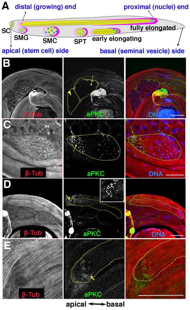
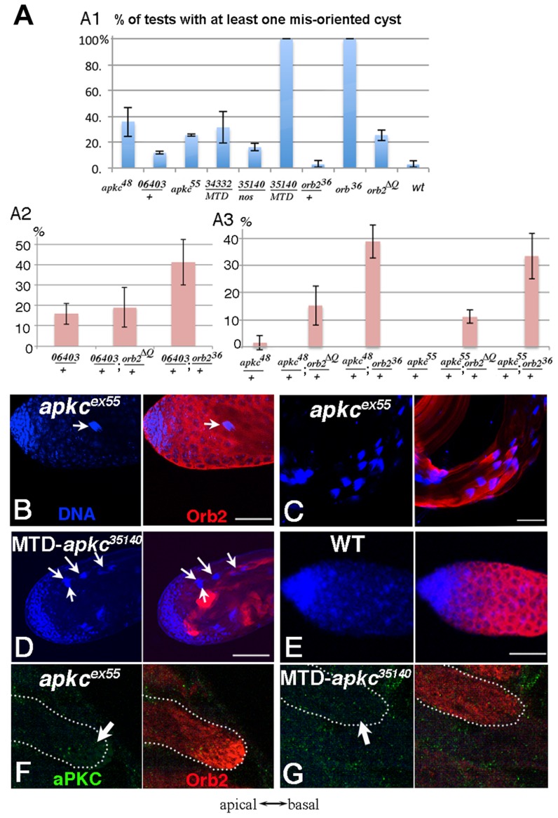
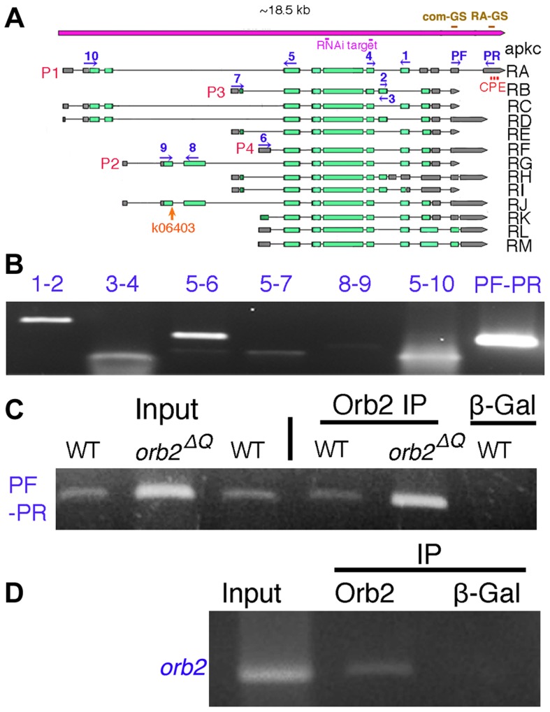
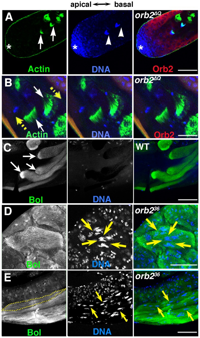
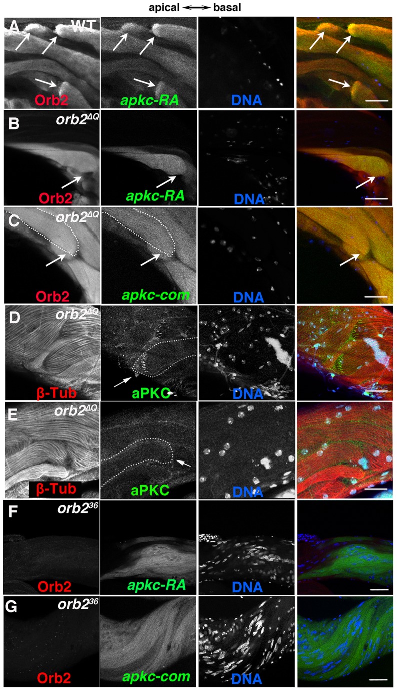
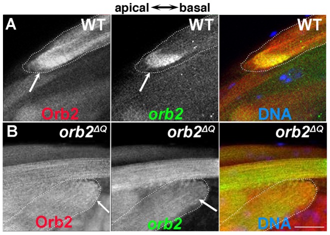
Mature Drosophila sperm are highly polarized cells--on one side is a nearly 2 mm long flagellar tail that comprises most of the cell, while on the other is the sperm head, which carries the gamete's genetic information. The polarization of the sperm cells commences after meiosis is complete and the 64-cell spermatid cyst begins the process of differentiation. The spermatid nuclei cluster to one side of the cyst, while the flagellar axonemes grows from the other. The elongating spermatid bundles are also polarized with respect to the main axis of the testis; the sperm heads are always oriented basally, while the growing tails extend apically. This orientation within the testes is important for transferring the mature sperm into the seminal vesicles. We show here that orienting cyst polarization with respect to the main axis of the testis depends upon atypical Protein Kinase C (aPKC), a factor implicated in polarity decisions in many different biological contexts. When apkc activity is compromised in the male germline, the direction of cyst polarization within this organ is randomized. Significantly, the mechanisms used to spatially restrict apkc activity to the apical side of the spermatid cyst are different from the canonical cross-regulatory interactions between this kinase and other cell polarity proteins that normally orchestrate polarization. We show that the asymmetric accumulation of aPKC protein in the cyst depends on an mRNA localization pathway that is regulated by the Drosophila CPEB protein Orb2. orb2 is required to properly localize and activate the translation of apkc mRNAs in polarizing spermatid cysts. We also show that orb2 functions not only in orienting cyst polarization with respect to the apical-basal axis of the testis, but also in the process of polarization itself. One of the orb2 targets in this process is its own mRNA. Moreover, the proper execution of this orb2 autoregulatory pathway depends upon apkc.


Conflict of interest statement

The authors have declared that no competing interests exist.

Figures

Figure 1
Figure 1. aPKC expression in spermatid cysts.
A) Stages of spermatogenesis. Germline cysts are in yellow, while the somatic cyst cells are labeled in dark pink (posterior or head cyst cell) and light pink (anterior or tail cyst cell) respectively. SC, stem cell; SMG, spermatogonia; SMC, spermatocyte; SPT, spermatid. B–E) aPKC expression during spermatogenesis. B) Localized aPKC is not observed in pre-meiotic (arrowhead) or meiotic (arrow) cysts. C) When the spermatid cyst polarizes, aPKC puncta (green) are clustered on the distal side of the polarizing cyst while the spermatid nuclei localize on the proximal side of the cyst. D, E) aPKC stripes at the tip of the growing flagellar axoneme (arrow). Insert in D is a zoom-in view of the marked window. All images are orientated with the apical end of the testes to the left and the basal to the right. Scale bar: 50 µm.
Figure 2
Figure 2. Spermatid cysts elongate in the wrong direction in apkc or orb2 mutants.
A) Frequency of testes that have at least one spermatid cyst polarized in the wrong orientation so that clustered spermatid cyst nuclei are found in the apical region instead of towards the base of the testis. apkc48 (apkcex48), 06403 (apkc06403) and apkc55 (apkcex55) are apkc mutant alleles. 34332 and 35140 are UAS-apkc RNAi lines (dsRNA targeting sites marked in Fig. 3A), while the Gal4 drivers are nos or MTD. MTD has three GAL4 drivers, pCOG-Gal4 which has an otu promoter, plus two different nos drivers, NGT-40 and nanos-Gal4. Error bars are standard deviation based on three experiments. Total number of testes scored for each genotype are (from left to right): Fig. A1: apkcex48: 36; apkc06403/+:33; apkcex55:27; MTD/UAS-apkc:34332:43; nos-GAL4/UAS-apkc:35140: 18; MTD/UAS-apkc:35140:16; orb36/+:55; orb36: 50; orb2ΔQ: 86; WT: 65. Fig. A2: apkc06403/+: 38; apkc06403/+; orb2ΔQ/+:40; apkc06403/+; orb236/+: 49; Fig. A3: apkcex48/+: 44; apkcex48/+; orb2ΔQ/+: 39; apkcex48/+; orb236/+: 39; apkcex55/+: 27; apkcex55/+; orb2ΔQ/+: 28; apkcex55/+; orb236/+: 37. B-D) Whole mount antibody staining of wild type or apkc mutant testes. Blue, DNA; red, Orb2. B) Spermatid nuclei clusters (arrow) are found in the spermatogonial region of the testis in apkc hypomorphic alleles. The example shown here is apkcex55. D) In the hypomorphic alleles, most spermatid nuclei clusters are located as in wild type on the basal side of testes. D) In MTD/UAS-apkc:35140 testes, multiple spermatid nuclei clusters are found in the apical region (arrows). (Not all spermatid cysts express Orb2 and/or are in focus). E) In wild type testes spermatid cyst nuclei are never seen at the apical end of the testis. F) aPKC (green) and Orb2 (red) in a misoriented elongating apkcex55 spermatid cyst. Note that the Orb2 comet head is present. G) aPKC (green) and Orb2 (red) in a misoriented elongating MTD/UAS-apkc:35140 testis. Note that the Orb2 comet is absent. This is also the case for correctly oriented MTD/UAS-apkc:35140 spermatid cysts (not shown). All images are orientated with apical side of the testes to the left and basal to the right. Scale bars: 50 µm.
Figure 3
Figure 3. apkc-RA and orb2 mRNAs associate with Orb2 in vivo.
A) The apkc transcription unit as annotated in http://flybase.org/reports/FBgn0261854.html. Transcripts expressed from four apkc promoters (P1–P4) plus a collection of alternatively spliced exons and UTRs are predicted to generate more than ten mRNAs. Green bar, protein coding exon; grey bar, UTR; solid black line, intron. apkc-RA CPE sites are labeled in red. Positions of primers used for RT-PCR experiments to identify different apkc mRNA species are marked in dark blue. The two dsRNA sequences used in the apkc RNAi knockdown experiments are marked in pink. Probes for the apkc Orb2 EMSAs, com-GS and RA-GS, are indicated above the gene in brown. B) apkc transcripts expressed in testes were detected by RT-PCR using the primer pairs as indicated. Not all of the primer pairs are specific to one of the four promoters, but some are specific to particular splice forms. C) apkc-RA mRNA can be immunoprecipitated with Orb2 antibodies from WT and orb2ΔQ testis extracts. β-Gal antibody (a negative control) didn't immunoprecipitate apkc-RA mRNA. D) Orb2 antibody immunoprecipitated orb2 mRNA from wild type testes while the control β-Gal antibody did not.
Figure 4
Figure 4. Localized accumulation of apkc mRNA, aPKC and Orb2 protein.
A, B) Testes were probed with apkc-com or apkc-RA specific fluorescent oligos. Panels highlight apkc expression at different stages of spermatogenesis. A) Left panel: mRNAs complementary to the apkc-com mRNA probe (specific for an exon that is present in all of the predicted apkc mRNA species – “bulk” apkc mRNA) were detected in the stem cell/spermatogonial/early spermatocytes region of the testis. Middle and right panel: “Bulk” apkc mRNA complementary to the apkc-com probe is also present at moderate levels in spermatocytes (SMC) and spermatids (SPT) (white arrows). In elongating spermatid cysts bulk apkc mRNA is localized in a comet pattern (yellow arrow). B) Left panel: Unlike “bulk” apkc mRNA, apkc-RA is not expressed in stem cells or spermatogonial cysts. Middle panel: apkc-RA is also absent in spermatocytes (SMC, arrowhead). Low levels of apkc-RA are first detected in spermatid cysts (SPT, white arrow) after meiosis is complete but prior to cyst polarization. Right panel: High levels of apkc-RA are present during the elongation phase (yellow arrow). At this stage apkc-RA accumulates in the characteristic comet pattern. C) aPKC protein stripes are found at the proximal (away from the elongating frontier) edge of the apkc-RA comet head. D) “Bulk” apkc mRNAs (detected with the apkc-com probe) and E) apkc-RA mRNAs co-localize with Orb2 in a comet pattern. The most extensive co-localization of mRNA and protein appears to be in the comet head. F) aPKC protein stripes accumulate at the proximal edge of the Orb2 protein comet head. All images are orientated with the apical end of the testis is to the left and basal end to the right. Scale bar: 20 µm.
Figure 5
Figure 5. orb2 is required for cyst polarization.
A and B) Green, phalloidin labeled Actin; Blue, DNA; Red, Orb2. A) Clustered nuclei of spermatid cysts (arrowheads) that have completed elongation and just assembled individualization complexes (ICs) (arrows) are found in the spermatogonial/early spermatocyte region of orb2ΔQ testis. B) White arrows point to two ICs in an orb2ΔQ testis that are moving in opposite directions in the middle of the testis (yellow arrows indicate directions of motion). C–E) Green, Bol; blue, DNA. C) Like Orb2, the translation factor Bol is localized a comet pattern during flagellar axoneme elongation in wild type testes . D) A bi-polar orb236 spermatid cyst that has Bol concentrated at both elongating ends, while the nuclei are in the middle of the cyst (arrows). E) A partially elongated orb236 spermatid cyst in which spermatid nuclei and Bol protein are scattered randomly. Dotted line outlines one among four spermatid cysts in the figure. Yellow arrows point out a few of the scattered nuclei. All images are orientated with apical side of the testes to the left and basal to the right. Scale bar: 50 µm.
Figure 6
Figure 6. orb2 is required for polarized accumulation of apkc-RA mRNA and aPKC protein in spermatid cysts.
A) In wild type testes, Orb2 and apkc-RA accumulate in a comet pattern in elongating spermatid cysts located towards the apical side of the testis. B and C) In orb2ΔQ spermatid cysts that elongate in the incorrect direction, Orb2ΔQ protein and both apkc-RA mRNA (detected with the apkc-RA probe, B) and “bulk” apkc mRNA (detected with the apkc-com probe, C) are distributed randomly instead of localizing in the comet pattern. Arrows in A indicate comet heads. In B and C the arrows indicate the expected position of the comet head. In the merged images on the far right, Red: Orb2; Green: apkc mRNA; Blue: DNA. D & E) aPKC protein (arrow) in orb2ΔQ spermatids elongating in the correct (D) or incorrect (E) orientation. orb2ΔQ spermatids elongating in the incorrect direction lack aPKC protein stripes at the tip of the elongating flagellar axonemes (compare arrows in D and E). In the merged images on the far right, Red: Tublin; Green: aPKC; Blue: DNA. F & G) apkc-RA (detected with the apkc-RA probe) and “bulk” apkc mRNAs (detected with the apkc-com probe) are expressed in orb236 spermatids but are not localized. In the merged image on right, Red: Orb2, Green: apkc-RA or bulk (apkc-com) mRNA, and Blue: DNA. All images are orientated with apical side of the testes to the left and basal to the right. Scale bar in A–D: 20 µm; in E and F: 50 µm.
Figure 7
Figure 7. orb2 autoregulates the localization and translation of orb2 mRNA.
A) Wild type spermatid cyst showing that Orb2 protein (red) and orb2 mRNA (green) are distributed in the characteristic comet pattern and display extensive co-localization, particularly in the comet head. B) In orb2ΔQ spermatids that elongate in the incorrect direction, Orb2ΔQ protein (red) and orb2ΔQ mRNA (green) are distributed uniformly in the cyst and there is no evidence of a comet head (expected position is indicated by arrow). All images are orientated with apical to the left and basal to the right. Scale bar: 20 µm.

Similar articles

Cited by

References

    1. Munro E, Bowerman B (2009) Cellular symmetry breaking during Caenorhabditis elegans development. Cold Spring Harb Perspect Biol 1: a003400. - PMC - PubMed
    1. Etemad-Moghadam B, Guo S, Kemphues KJ (1995) Asymmetrically distributed PAR-3 protein contributes to cell polarity and spindle alignment in early C. elegans embryos. Cell 83: 743–752. - PubMed
    1. Watts JL, Etemad-Moghadam B, Guo S, Boyd L, Draper BW, et al. (1996) par-6, a gene involved in the establishment of asymmetry in early C. elegans embryos, mediates the asymmetric localization of PAR-3. Development 122: 3133–3140. - PubMed
    1. Tabuse Y, Izumi Y, Piano F, Kemphues KJ, Miwa J, et al. (1998) Atypical protein kinase C cooperates with PAR-3 to establish embryonic polarity in Caenorhabditis elegans. Development 125: 3607–3614. - PubMed
    1. Hao Y, Boyd L, Seydoux G (2006) Stabilization of cell polarity by the C. elegans RING protein PAR-2. Dev Cell 10: 199–208. - PMC - PubMed

Publication types