

Circadian variation in vascular function and regenerative capacity in healthy humans
- PMID: 24830296
- PMCID: PMC4309078
- DOI: 10.1161/JAHA.114.000845
Circadian variation in vascular function and regenerative capacity in healthy humans
Abstract
Background: Progenitor cells (PCs) are mobilized in response to vascular injury to effect regeneration and repair. Recruitment of PCs requires intact nitric oxide (NO) synthesis by endothelial cells, and their number and activity correlate with cardiovascular disease risk burden and future outcomes. Whereas cardiovascular vulnerability exhibits a robust circadian rhythm, the 24-hour variation of PCs and their inter-relation with vascular function remain unknown. We investigated the circadian variation of PCs and vascular function with the hypothesis that this will parallel the pattern observed for cardiovascular events (CVEs).
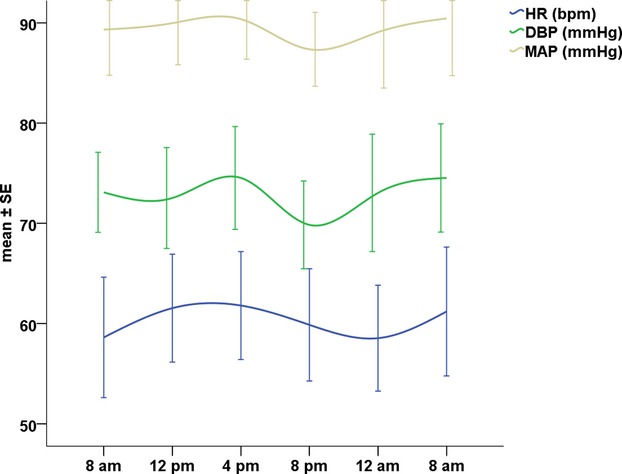
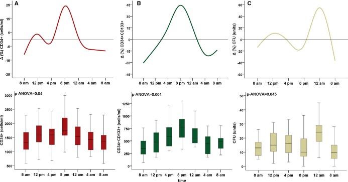
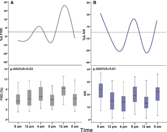
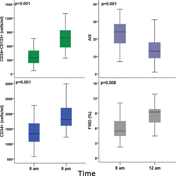
Methods and results: In 15 healthy subjects (9 men, 37±16 years), circulating PCs and vascular function were measured at 8 am, noon, 4 pm, 8 pm, midnight, 4 am (only PCs counts), and 8 am the following day. Circulating PCs were enumerated as mononuclear cells (MNCs; CD45(med)) that express CD34 as well as CD133, and their activity was assessed as the number of colonies formed by culturing MNCs. Vascular function was evaluated by measurement of endothelium-dependent, flow-mediated vasodilation (FMD) of the brachial artery and tonometry-derived indices of arterial stiffness. Higher CD34(+) and CD34(+)/CD133(+) cell counts were observed at 8 pm than any other time of the day (P-ANOVA=0.038 and <0.001; respectively) and were lowest at 8 am. PC colony formation was highest at midnight (P-ANOVA=0.045) and lowest in the morning hours. FMD was highest at midnight and lowest at 8 am and 8 pm, and systemic arterial stiffness was greatest at 8 am and lowest at 4 pm and midnight (P-ANOVA=0.03 and 0.01; respectively).
Conclusion: A robust circadian variation in PC counts and vascular function occurs in healthy humans and both exhibit an unfavorable profile in the morning hours that parallels the preponderance of CVEs at these times. Whether these changes are precipitated by awakening and time-dependent physical activity or governed by the endogenous circadian clock needs to be further investigated.
Keywords: arterial stiffness; circadian variation; endothelial function; progenitor cells.
© 2014 The Authors. Published on behalf of the American Heart Association, Inc., by Wiley Blackwell.
Figures
Similar articles
-
Sex Differences in Circulating Progenitor Cells.J Am Heart Assoc. 2017 Oct 3;6(10):e006245. doi: 10.1161/JAHA.117.006245. J Am Heart Assoc. 2017. PMID: 28974500 Free PMC article.
-
Early morning attenuation of endothelial function in healthy humans.Circulation. 2004 Jun 1;109(21):2507-10. doi: 10.1161/01.CIR.0000128207.26863.C4. Epub 2004 May 10. Circulation. 2004. PMID: 15136499
-
Age and Human Regenerative Capacity Impact of Cardiovascular Risk Factors.Circ Res. 2016 Sep 16;119(7):801-9. doi: 10.1161/CIRCRESAHA.116.308461. Epub 2016 Jul 19. Circ Res. 2016. PMID: 27436845 Free PMC article.
-
Endothelial progenitor cells in vascular repair and remodeling.Pharmacol Res. 2008 Aug;58(2):148-51. doi: 10.1016/j.phrs.2008.07.008. Epub 2008 Aug 5. Pharmacol Res. 2008. PMID: 18722530 Review.
-
Endothelial progenitor cells and vascular repair.Curr Opin Hematol. 2014 May;21(3):224-8. doi: 10.1097/MOH.0000000000000041. Curr Opin Hematol. 2014. PMID: 24637956 Free PMC article. Review.
Cited by
-
Tumor necrosis factor-alpha antagonism with etanercept improves endothelial progenitor cell counts in patients with psoriasis: etanercept, vascular function and endothelial progenitor cells in psoriasis.Int J Cardiol. 2015 Mar 1;182:387-9. doi: 10.1016/j.ijcard.2014.12.093. Epub 2014 Dec 27. Int J Cardiol. 2015. PMID: 25617605 Free PMC article. No abstract available.
-
Core circadian clock gene expression in human dental pulp-derived cells in response to L-mimosine, hypoxia and echinomycin.Eur J Oral Sci. 2018 Aug;126(4):263-271. doi: 10.1111/eos.12535. Eur J Oral Sci. 2018. PMID: 30006964 Free PMC article.
-
Insomnia Symptoms Are Associated With Abnormal Endothelial Function.J Cardiovasc Nurs. 2017 Jan/Feb;32(1):78-85. doi: 10.1097/JCN.0000000000000295. J Cardiovasc Nurs. 2017. PMID: 26488555 Free PMC article.
-
The association between baseline circulating progenitor cells and vascular function: The role of aging and risk factors.Vasc Med. 2022 Dec;27(6):532-541. doi: 10.1177/1358863X221116411. Epub 2022 Sep 5. Vasc Med. 2022. PMID: 36062298 Free PMC article.
-
Longitudinal timing of physical activity and associated cardiometabolic and behavioral health outcomes in young adults.Ann Behav Med. 2025 Jan 4;59(1):kaae084. doi: 10.1093/abm/kaae084. Ann Behav Med. 2025. PMID: 39658316
References
-
- Muller JE, Stone PH, Turi ZG, Rutherford JD, Czeisler CA, Parker C, Poole WK, Passamani E, Roberts R, Robertson T, Sobel BE, Willerson JT, Braunwald E. Circadian variation in the frequency of onset of acute myocardial infarction. N Engl J Med. 1985; 313:1315-1322. - PubMed
-
- Mulcahy D, Keegan J, Cunningham D, Quyyumi A, Crean P, Park A, Wright C, Fox K. Circadian variation of total ischaemic burden and its alteration with anti‐anginal agents. Lancet. 1988; 2:755-759. - PubMed
-
- Marler JR, Price TR, Clark GL, Muller JE, Robertson T, Mohr JP, Hier DB, Wolf PA, Caplan LR, Foulkes MA. Morning increase in onset of ischemic stroke. Stroke. 1989; 20:473-476. - PubMed
-
- Willich SN, Levy D, Rocco MB, Tofler GH, Stone PH, Muller JE. Circadian variation in the incidence of sudden cardiac death in the Framingham heart study population. Am J Cardiol. 1987; 60:801-806. - PubMed
-
- Panza JA, Epstein SE, Quyyumi AA. Circadian variation in vascular tone and its relation to alpha‐sympathetic vasoconstrictor activity. N Engl J Med. 1991; 325:986-990. - PubMed
Publication types
MeSH terms
Grants and funding
LinkOut - more resources
Full Text Sources
Other Literature Sources
Research Materials
Miscellaneous
