Review
. 2014 Jun;14(6):377-91.
doi: 10.1038/nri3667. Epub 2014 May 16.

Positive and negative selection of the T cell repertoire: what thymocytes see (and don't see)

Affiliations
Review

Positive and negative selection of the T cell repertoire: what thymocytes see (and don't see)

Ludger Klein et al. Nat Rev Immunol. 2014 Jun.

Abstract

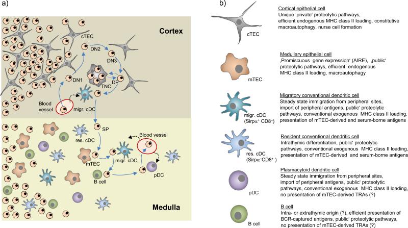
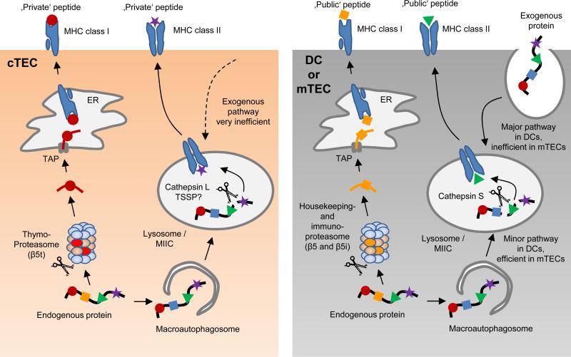
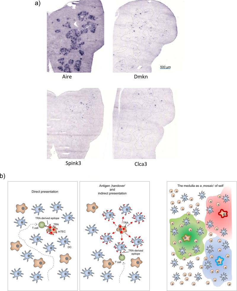
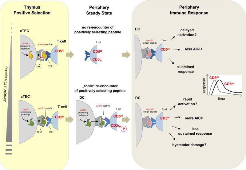
The fate of developing T cells is specified by the interaction of their antigen receptors with self-peptide-MHC complexes that are displayed by thymic antigen-presenting cells (APCs). Various subsets of thymic APCs are strategically positioned in particular thymic microenvironments and they coordinate the selection of a functional and self-tolerant T cell repertoire. In this Review, we discuss the different strategies that these APCs use to sample and process self antigens and to thereby generate partly unique, 'idiosyncratic' peptide-MHC ligandomes. We discuss how the particular composition of the peptide-MHC ligandomes that are presented by specific APC subsets not only shapes the T cell repertoire in the thymus but may also indelibly imprint the behaviour of mature T cells in the periphery.


Figures

Figure 1
Figure 1. Stromal cell interactions during T cell development
(a) Successive stages of double-negative (DN) T cell development are accompanied by an outward movement of thymocytes towards the sub-capsular zone. Subsequent to β-selection at the DN3 stage, double-positive (DP) cells ‘randomly walk’ through the outer cortex, which possibly facilitates the ‘scanning’ of cortical thymic epithelial cells (cTECs) for positively selecting ligands. At this stage, DP thymocytes may be engulfed by cTECs and form so-called thymic nurse cells (TNCs), whereby the molecular control and physiological relevance of this process remains to be established. Interactions of DP cells with cortical conventional dendritic cells (cDCs) may lead to negative selection. It remains open whether these cortical cDCs exclusively belong to the migratory Sirpα+ subset. Positively selected, CD4 or CD8 lineage-committed thymocytes relocate into the medulla by directed migration. Upon reaching the medulla, single-positive (SP) cells again assume a ‘random walk’ motion pattern. Through this random migration, SP cells may now ‘scan’ resident (res.) and migratory (migr.) cDCs, medullary thymic epithelial cells (mTECs), plasmacytoid dendritic cells (pDCs) and B cells. It is estimated that SP cells engage in around five contacts with antigen presenting cells (APCs) per hour, so that over their 4-5 days residency in the medulla, T cells may serially interact with several hundred APCs. (b) Key functional properties of thymic APCs discussed in this Review.
Figure 2
Figure 2. Unique proteolytic pathways generate ‘private’ MHC-bound peptides in cTECs
Processing of a given endogenous protein substrate by cTECs may give rise to unique, ‘private’ peptides, which differ from ‘public’ peptides generated by mTECs and DCs. MHC class I-bound peptides on the surface of cortical thymic epithelial cells (cTECs) are predominantly processed by proteasomes containing the catalytic subunit β5t (so called thymoproteasomes). Due to a distinct proteolytic activity of the thymoproteasome, this is likely to lead to the generation of cTEC-specific, ‘private’ peptide epitopes that differ from ‘public’ epitopes generated by mTECs or DCs through the housekeeping proteasome or the immuno-proteasome. MHC class II-bound peptides on cTECs seem to be mostly derived from an unconventional, endogenous MHC class II-loading pathway that involves the macroautophagy-mediated shuttling of cytoplasmic proteins into lysosomes. In this proteolytic compartment, processing by the proteases cathepsin L and thymus-specific serin protease (TSSP) may generate unique ‘private’ peptides. MHC class II-bound peptides on mTECs may likewise be mostly derived from macroautophagy–mediated endogenous MHC class II-loading; however, the lysosomal proteases that generate MHC class II-bound peptides in mTECs differ from those in cTECs, being essentially identical to those used by DCs for the processing of exogenously-derived substrates along the ‘conventional’, exogenous MHC class II pathway. Of note, it is likely that the pMHC ligandome of cTECs represents a mixture of ‘private’ and ‘public’ peptides that are uniquely present on cTECs or shared with other APCs, respectively (see Figure 4).
Figure 3
Figure 3. Topological aspects of ‘promiscuous gene expression’ and direct versus indirect presentation of TRAs
(a) Ttissue-restricted antigens (TRAs) are expressed by only a small subset of mTECs at any given point in time: In-situ hybridization of a section through an entire thymic lobe for Aire mRNA expression (left) shows that Aire-expressing cells are densely packed in medullary regions. By contrast, transcripts of three representative TRAs (dermakine, Dmkn; Serin protease inhibitor kazal type 3, Spink3; chloride channel calcium activated 3; Clca3) are only detectable in very few cells that are scattered throughout medullary areas (dotted lines). (b) Tolerogenic presentation of TRAs that are expressed by few mTECs may occur in two not mutually exclusive ways: (left) direct presentation by TRA-expressing mTECs themselves, whereby efficient endogenous MHC class II-loading by mTECs in conjunction with serial ‘scanning’ of multiple medullary APCs by thymocytes increases the likelihood of cognate self-antigen interactions. (middle) ‘Antigen handover’ to neighbouring cDCs may extend the area of tolerogenic presentation in a mosaic fashion beyond the topologically restricted expression pattern (right). The mechanistic details of this ‘directional antigen transfer’ transfer remain to be established. It is conceivable that TRAs are released or shed in soluble form to be subsequently captured and processed by cDCs for presentation on MHC class I or II. Apoptosis of terminally differentiated mTECs may lead to the release of apoptotic fragments that can also transfer mTEC-derived self-antigens to cDCs. In addition, functional peptide-MHC ligands are unidirectionally translocated from mTECs to DCs , .
Figure 4
Figure 4. Consequences of positive selection by ‘private’ versus ‘public‘ peptides: a hypothesis
(Upper panel) ‘Private’ peptides generated through unique proteolytic pathways in cortical thymic epithelial cells (cTECs) may preferentially support selection of CD5low T cell clones via interactions at the lower end of the affinity range that is permissive for positive selection. One determinant of these ‘low strength’ interactions could be that private peptides are weak MHC binders, indicated here by the loose fit between peptide and MHC (red arrow). In the periphery, T cells selected in this way do not re-encounter the positively selecting peptides and hence do not receive tonic signals. As a consequence, their CD3ς chains are not pre-loaded with basal phosphorylation. Yet, it remains possible that CD5low clones receive a degree of tonic input through exposure to cross-reactive ‘public’ peptides in the periphery. (Lower panel) Public peptides may preferentially support selection of CD5hi clones via positively selecting interactions at the relatively higher end of the affinity range. Public peptides might be good MHC binders that generate ‘low strength’ interactions by loosely binding to the TCR (red arrow). In the periphery, continual interactions with the very same peptides support T cell homeostasis and mediate partial CD3ς chain phosphorylation. During an immune response to foreign antigens, CD5low and CD5hi T cell clones of identical specificity may differentially respond with respect to timing and magnitude of clonal expansion and contraction. The dominance of either type of responder might vary with parameters such as duration and anatomical distribution of the infection.

Comment in

References

    1. Kyewski B, Klein L. A central role for central tolerance. Annual review of immunology. 2006;24:571–606. - PubMed
    1. Nakagawa Y, Ohigashi I, Nitta T, Sakata M, Tanaka K, et al. Thymic nurse cells provide microenvironment for secondary T cell receptor alpha rearrangement in cortical thymocytes. Proceedings of the National Academy of Sciences of the United States of America. 2012;109:20572–20577. - PMC - PubMed
    1. Klein L, Hinterberger M, Wirnsberger G, Kyewski B. Antigen presentation in the thymus for positive selection and central tolerance induction. Nature reviews. Immunology. 2009;9:833–844. - PubMed
    1. Florea BI, Verdoes M, Li N, van der Linden WA, Geurink PP, et al. Activity-based profiling reveals reactivity of the murine thymoproteasome-specific subunit beta5t. Chemistry & biology. 2010;17:795–801. - PMC - PubMed
    1. Murata S, Sasaki K, Kishimoto T, Niwa S, Hayashi H, et al. Regulation of CD8+ T cell development by thymus-specific proteasomes. Science. 2007;316:1349–1353. - PubMed

Publication types