. 2014 May 30;5(10):3273-86.
doi: 10.18632/oncotarget.1926.

Targeting human apurinic/apyrimidinic endonuclease 1 (APE1) in phosphatase and tensin homolog (PTEN) deficient melanoma cells for personalized therapy

Affiliations

Targeting human apurinic/apyrimidinic endonuclease 1 (APE1) in phosphatase and tensin homolog (PTEN) deficient melanoma cells for personalized therapy

Rachel Abbotts et al. Oncotarget. .

Abstract

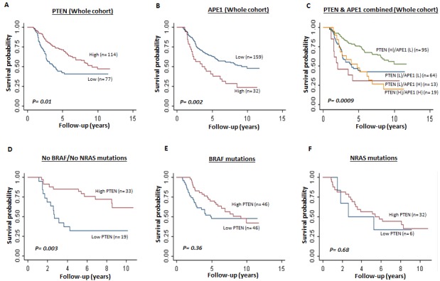
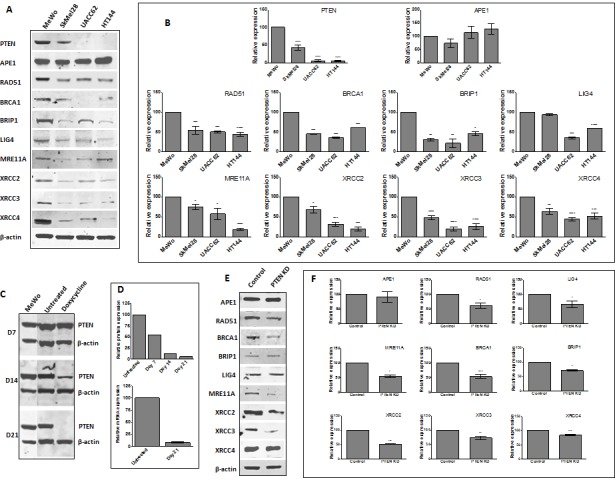
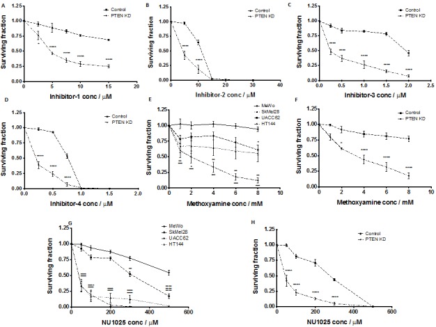
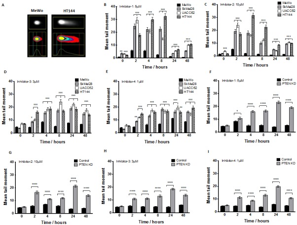
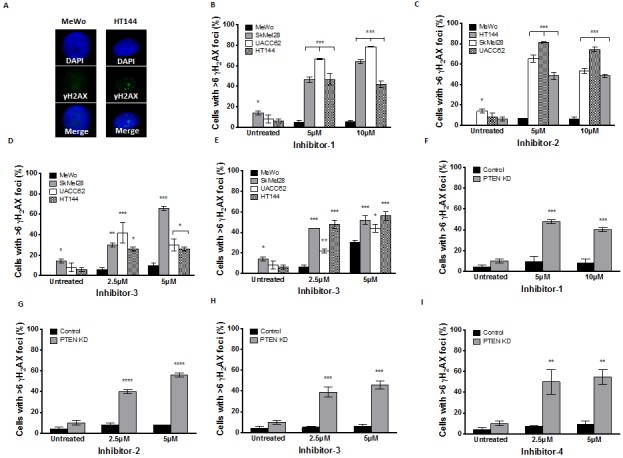
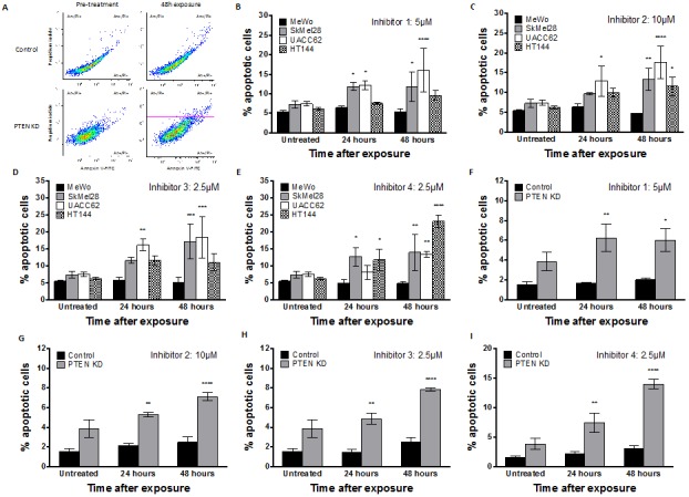
Phosphatase and tensin homolog (PTEN) loss is associated with genomic instability. APE1 is a key player in DNA base excision repair (BER) and an emerging drug target in cancer. We have developed small molecule inhibitors against APE1 repair nuclease activity. In the current study we explored a synthetic lethal relationship between PTEN and APE1 in melanoma. Clinicopathological significance of PTEN mRNA and APE1 mRNA expression was investigated in 191 human melanomas. Preclinically, PTEN-deficient BRAF-mutated (UACC62, HT144, and SKMel28), PTEN-proficient BRAF-wildtype (MeWo), and doxycycline-inducible PTEN-knockout BRAF-wildtype MeWo melanoma cells were DNA repair expression profiled and investigated for synthetic lethality using a panel of four prototypical APE1 inhibitors. In human tumours, low PTEN mRNA and high APE1 mRNA was significantly associated with reduced relapse free and overall survival. Pre-clinically, compared to PTEN-proficient cells, PTEN-deficient cells displayed impaired expression of genes involved in DNA double strand break (DSB) repair. Synthetic lethality in PTEN-deficient cells was evidenced by increased sensitivity, accumulation of DSBs and induction of apoptosis following treatment with APE1 inhibitors. We conclude that PTEN deficiency is not only a promising biomarker in melanoma, but can also be targeted by a synthetic lethality strategy using inhibitors of BER, such as those targeting APE1.


Conflict of interest statement

The authors disclose no potential conflicts of interest

Figures

Figure 1
Figure 1. Kaplan Meier curves showing overall survival in melanoma
A. Whole cohort (PTEN mRNA high and low, p=0.01). B. Whole cohort (APE1 mRNA high and low, p=0.002). C. Whole cohort (PTEN and APE1 m RNA combined high and low, p=0.0009). D. Tumours with no BRAF/NRAS mutations (PTEN mRNA high and low, p=0.003). E. Tumours with BRAF V600E mutation (PTEN mRNA high and low, p=0.36). F. Tumours with NRAS mutations (PTEN mRNA high and low, p=0.68).
Figure 2
Figure 2
A. Representative Western blots of PTEN and selected DNA repair proteins in human melanoma cell lines. B. Quantification of PTEN and selected DNA repair factor protein levels in melanoma cell lines. C. Generation of doxycycline-inducible PTEN knockdown MeWo cells. Following lentiviral transduction, continuous doxycycline exposure induced PTEN shRNA expression. Whole cell lysates were examined for PTEN protein levels every 7 days, with almost complete absence observed by day 21. This was confirmed by mRNA analysis by qRT-PCR (D). E. Representative Western blots and quantification (F) of PTEN and selected DNA repair proteins in control and PTEN knockdown (KD) cell line. * p<0.05, ** p<0.01, *** p<0.001, **** p<0.0001 compared to control cells.
Figure 3
Figure 3. Aldehyde reactive probe assay confirming target inhibition in MeWo, SkMel28, UACC62 and HT144 cells treated with inhibitor -1
(A), inhibitor -2 (B), inhibitor -3 (C) and inhibitor -4 (D) Clonogenic survival assays in MeWo, SkMel28, UACC62 and HT144 cells treated with inhibitor-1 (E), inhibitor-2 (F), inhibitor-3 (G) and inhibitor-4 (H). Inhibitors were added at the indicated concentrations (see methods for details). * p<0.05, ** p<0.01, *** p<0.001, **** p<0.0001 compared to MeWo (PTEN-wildtype).
Figure 4
Figure 4. Clonogenic survival assays in control MeWo cells and PTEN knockdown (KD) MeWo cells treated with inhibitor-1
(A), inhibitor-2 (B), inhibitor-3 (C) and inhibitor-4 (D) Clonogenic survival assays in MeWo, SkMel28, UACC62 and HT144 cells treated with methoxyamine (E). Clonogenic survival assays in control MeWo cells and PTEN knockdown (KD) MeWo cells treated with methoxyamine (F). Clonogenic survival assays in MeWo, SkMel28, UACC62 and HT144 cells treated with NU1025 (G). Clonogenic survival assays in control MeWo cells and PTEN knockdown (KD) MeWo cells treated with NU1025 (H). Inhibitors were added at the indicated concentrations (see methods for details). * p<0.05, ** p<0.01, *** p<0.001, **** p<0.0001 compared to MeWo (PTEN-wildtype).
Figure 5
Figure 5. Neutral comet assay was performed at various time points after APE1 inhibitor exposure as indicated in methods
A. Example of increased DNA damage comet ‘tail’ in PTEN-deficient HT144 compared to MeWo cells following 24 hours exposure to inhibitor-1. Significantly increased mean tail moment was observed in PTEN-deficient SkMel28, UACC62 and HT144 cells compared to MeWo cells following treatment with inhibitor-1 (B), inhibitor-2 (C), inhibitor 3 (D) and inhibitor 4 (E). Similarly, significantly increased mean tail moment was observed in PTEN knockdown (KD) MeWo cells compared to control MeWo cells treated with inhibitor-1 (F), inhibitor-2 (G), inhibitor 3 (H) and inhibitor 4 (I). ** p<0.01, *** p<0.001, compared to MeWo (PTEN-wildtype).
Figure 6
Figure 6
A. γH2AX immunocytochemistry following 24 hours inhibitor-1 exposure demonstrates increased foci in PTEN-deficient HT144 cells compared to PTEN-wildtype MeWo. Significantly increased γH2AX foci was observed in PTEN-deficient SkMel28, UACC62 and HT144 cells compared to MeWo cells following 24 hours treatment with inhibitor-1 (B), inhibitor-2 (C), inhibitor-3 (D) or inhibitor-4 (E). Similar significantly increased γH2AX foci was observed in PTEN knockdown (KD) MeWo cells compared to control MeWo cells following 24 hours treatment with inhibitor-1 (F), inhibitor-2 (G), inhibitor-3 (H) or inhibitor-4 (I). * p<0.05, ** p<0.01, *** p<0.001, compared to MeWo (PTEN-wildtype).
Figure 7
Figure 7
A. Apoptosis detection by FITC-annexin V flow cytometric analysis demonstrates an increased apoptotic fraction in HT144 following 48 hours exposure to inhibitor-1, as observed by increased FITC-Annexin V staining in upper and lower right quadrants. Significantly increased apoptotic fraction was observed in PTEN-deficient SkMel28, UACC62 and HT144 cells compared to MeWo cells following 48 hours treatment with indicated concentrations of inhibitor-1 (B), inhibitor-2 (C), inhibitor-3 (D) or inhibitor-4 (E). Similar significantly increased apoptotic fraction was observed in PTEN knockdown (KD) MeWo cells compared to control MeWo cells following 48 hours treatment with inhibitor-1 (F), inhibitor-2 (G), inhibitor-3 (H) or inhibitor-4 (I). * p<0.05, ** p<0.01, *** p<0.001, compared to MeWo (PTEN-wildtype).

References

    1. Abbotts R, Madhusudan S. Human AP endonuclease 1 (APE1): from mechanistic insights to druggable target in cancer. Cancer Treat Rev. 2010;36(5):425–435. - PubMed
    1. Dianov GL, Sleeth KM, Dianova, Allinson SL. Repair of abasic sites in DNA. Mutat Res. 2003;531(1-2):157–163. - PubMed
    1. Li M, Wilson DM. Human Apurinic/Apyrimidinic Endonuclease 1. Antioxid Redox Signal. (3rd) 2014;20(4):678–707. - PMC - PubMed
    1. Evans AR, Limp-Foster M, Kelley MR. Going APE over ref-1. Mutat Res. 2000;461(2):83–108. - PubMed
    1. Wang D, Xiang DB, Yang XQ, Chen LS, Li MX, Zhong ZY, Zhang YS. APE1 overexpression is associated with cisplatin resistance in non-small cell lung cancer and targeted inhibition of APE1 enhances the activity of cisplatin in A549 cells. Lung Cancer. 2009;66(3):298–304. - PubMed

Publication types

MeSH terms