. 2014 Jun 3;111(22):8155-60.
doi: 10.1073/pnas.1321502111. Epub 2014 May 15.

The structural analysis of shark IgNAR antibodies reveals evolutionary principles of immunoglobulins

Affiliations

The structural analysis of shark IgNAR antibodies reveals evolutionary principles of immunoglobulins

Matthias J Feige et al. Proc Natl Acad Sci U S A. .

Abstract

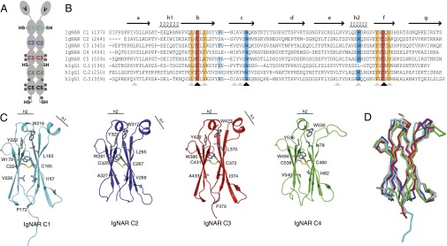
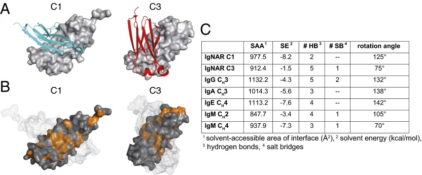
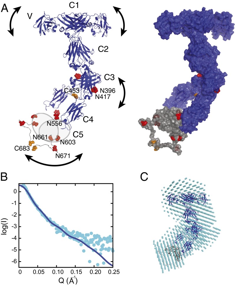
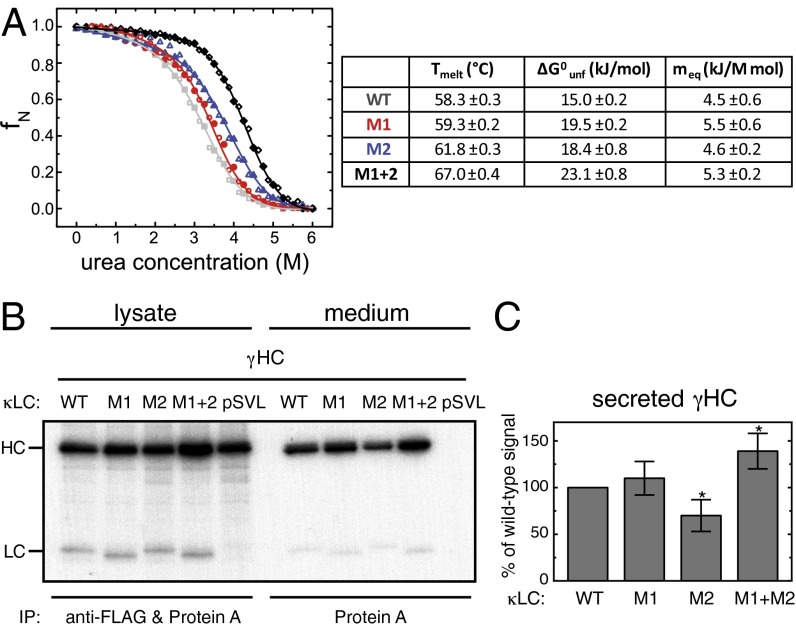
Sharks and other cartilaginous fish are the phylogenetically oldest living organisms that rely on antibodies as part of their adaptive immune system. They produce the immunoglobulin new antigen receptor (IgNAR), a homodimeric heavy chain-only antibody, as a major part of their humoral adaptive immune response. Here, we report the atomic resolution structure of the IgNAR constant domains and a structural model of this heavy chain-only antibody. We find that despite low sequence conservation, the basic Ig fold of modern antibodies is already present in the evolutionary ancient shark IgNAR domains, highlighting key structural determinants of the ubiquitous Ig fold. In contrast, structural differences between human and shark antibody domains explain the high stability of several IgNAR domains and allowed us to engineer human antibodies for increased stability and secretion efficiency. We identified two constant domains, C1 and C3, that act as dimerization modules within IgNAR. Together with the individual domain structures and small-angle X-ray scattering, this allowed us to develop a structural model of the complete IgNAR molecule. Its constant region exhibits an elongated shape with flexibility and a characteristic kink in the middle. Despite the lack of a canonical hinge region, the variable domains are spaced appropriately wide for binding to multiple antigens. Thus, the shark IgNAR domains already display the well-known Ig fold, but apart from that, this heavy chain-only antibody employs unique ways for dimerization and positioning of functional modules.

Keywords: antibody structure; protein engineering; protein evolution; protein folding.


Conflict of interest statement

Conflict of interest statement: A patent for optimized antibodies based on the results presented in this study has been filed.

Figures

Fig. 1.
Fig. 1.
Sequence and structure of IgNAR domains C1–C4. (A) Schematic of the secreted dimeric IgNAR molecule, comprising one variable (V) and five constant (C1–C5) domains. Predicted glycosylation sites are shown as gray hexagons. Cysteines that are not part of the intradomain disulfide bridges are indicated (–SH). The secretory tail is C terminally of the C5 domain. (B) Sequence alignment of IgNAR C1–C5 with the human IgG1 HC domains CH1–CH3. Conserved cysteines are highlighted in red, and conserved hydrophobic residues of a YxCxY (Y, hydrophobic residue) motif around the disulfide bridge are highlighted in orange. Conserved tryptophans in strand c and the second helix are highlighted in blue, and the cis-proline residue in the loop between strand b and c is depicted in cyan. Secondary structure elements are indicated above the alignment. Black arrows indicate strictly conserved residues, and gray arrows homologous residues. (C) Ribbon diagram of the isolated constant IgNAR domains C1–C4 (C1, cyan; C2, blue; C3, red; C4, green; colors like in A). Residues marked in the alignment are shown in stick representation, the small helices are indicated. (D) Superposition of the IgNAR C1-4 domains (C1, cyan; C2, blue; C3, red; C4, green) on a human IgG CH3 domain (gray, Protein Data Bank ID code 1HZH).
Fig. 2.
Fig. 2.
Characterization of the IgNAR C1 and C3 dimerization interfaces. (A) Ribbon and surface diagram of the IgNAR C1 and C3 dimers. The two subunits are colored in cyan and gray (C1) or red and gray (C3), respectively. (B) Hydrophobic residues within the C1 and C3 dimerization interfaces are shown in orange. One monomer is in surface representation; its counterpart is shown as a mesh surface. (C) Comparison of the dimerization interfaces of IgNAR C1 and C3 and different human and murine dimeric domains, as determined by the PISA server (48) (Protein Data Bank ID codes IgG CH3, 3HKF; IgA CH3, 1OW0; IgE CH4, 1O0V; IgM CH2, 4JVU; and IgM CH4, 4JVW).
Fig. 3.
Fig. 3.
A model of the complete IgNAR antibody. (A) A structural model of full-length IgNAR based on restraints from the C1 and C3 dimer structures and intermolecular disulfide bonds, derived by docking with HADDOCK (26). (Left) Crystal structures of the variable domain (Protein Data Bank ID code 1SQ2) and constant domains C1–C4. Because of the lack of structural information for C5, it is drawn as a gray oval. Cysteines forming interchain disulfide bonds are highlighted in orange and predicted glycosylation sites in red, respectively. The arced arrows indicate flexibility within the molecule. (Right) The same model is shown in surface view, including a possible conformation of the flexible C4–C5, as adopted in the lowest energy structure of the best cluster, assessed by the HADDOCK score and SAXS χ2. (B) SAXS data of full-length IgNAR (cyan dots). The Q range above 0.25 Å−1 has been removed because of noise. The blue line corresponds to the back-calculated curve from the HADDOCK-derived structure with the best fit to the SAXS data (shown in A; χ2 = 1.43). (C) An ab initio bead model derived from the SAXS data, using DAMMIF (27), superimposed with the lowest-energy structure of the HADDOCK cluster with the best fit to the SAXS data, using SUPCOMB (49).
Fig. 4.
Fig. 4.
Stability and oligomerization state of the constant IgNAR domains. C1–C4 were reversibly unfolded by guanidinium chloride (GdmCl) (C1, cyan; C2, blue; C3, red; C4, green; unfolding, closed circles; refolding, open circles). Midpoints of thermal transitions and thermodynamic parameters obtained by GdmCl-induced unfolding transitions are listed in the table (Tmelt, melting temperature; ΔG0unf, free energy of unfolding; meq, cooperativity parameter). The association state and dissociation constant Kd of the domains in solution were obtained by analytical ultracentrifugation. All data are shown ± SD.
Fig. 5.
Fig. 5.
The effect of IgNAR-based mutations of a human CL domain on domain stability and antibody secretion. (A) Thermal stabilities and thermodynamic parameters derived from urea melts are shown for the wt CL domain and mutants M1, M2, and M1+2. Urea melts are shown on top (wt, gray; M1, red; M2, blue; M1+2, black; unfolding, closed circles; refolding, open circles). (B) LCWT, LCM1, LCM2, and LCM1+2 or empty vector (pSVL) were coexpressed with γHCs, as indicated. Cells were metabolically labeled for 1 h and either immediately lysed (lysate) or subsequently chased for 24 h before analysis of the medium (medium). Lysates were immunoprecipitated with anti-FLAG antibody to capture the LCs together with protein A to isolate the HCs; media were immunoprecipitated with protein A only to detect LC-induced HC secretion. (C) HC secretion data were quantified by phosphorimager analysis [n = 7 ± SD; data that are statistically significantly different from the wt (P ≤ 0.05) are marked with an asterisk].

References

    1. Cooper MD, Alder MN. The evolution of adaptive immune systems. Cell. 2006;124(4):815–822. - PubMed
    1. Flajnik MF, Kasahara M. Origin and evolution of the adaptive immune system: Genetic events and selective pressures. Nat Rev Genet. 2010;11(1):47–59. - PMC - PubMed
    1. Blair JE, Hedges SB. Molecular phylogeny and divergence times of deuterostome animals. Mol Biol Evol. 2005;22(11):2275–2284. - PubMed
    1. Dooley H, Flajnik MF. Antibody repertoire development in cartilaginous fish. Dev Comp Immunol. 2006;30(1-2):43–56. - PubMed
    1. England JL, Haran G. Role of solvation effects in protein denaturation: From thermodynamics to single molecules and back. Annu Rev Phys Chem. 2011;62:257–277. - PMC - PubMed

Publication types

MeSH terms