

Autophagy inhibition by sustained overproduction of IL6 contributes to arsenic carcinogenesis
- PMID: 24830721
- PMCID: PMC4102645
- DOI: 10.1158/0008-5472.CAN-13-3182
Autophagy inhibition by sustained overproduction of IL6 contributes to arsenic carcinogenesis
Abstract
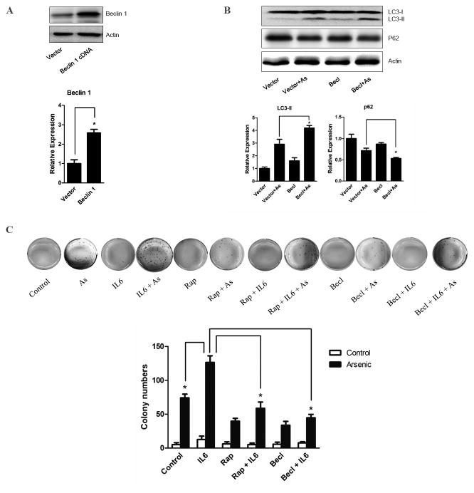
Chronic inflammation has been implicated as an etiologic factor in cancer, whereas autophagy may help preserve cancer cell survival but exert anti-inflammatory effects. How these phenomenas interact during carcinogenesis remains unclear. We explored this question in a human bronchial epithelial cell-based model of lung carcinogenesis that is mediated by subchronic exposure to arsenic. We found that sustained overexpression of the pro-inflammatory IL6 promoted arsenic-induced cell transformation by inhibiting autophagy. Conversely, strategies to enhance autophagy counteracted the effect of IL6 in the model. These findings were confirmed and extended in a mouse model of arsenic-induced lung cancer. Mechanistic investigations suggested that mTOR inhibition contributed to the activation of autophagy, whereas IL6 overexpression was sufficient to block autophagy by supporting Beclin-1/Mcl-1 interaction. Overall, our findings argued that chronic inflammatory states driven by IL6 could antagonize autophagic states that may help preserve cancer cell survival and promote malignant progression, suggesting a need to uncouple inflammation and autophagy controls to enable tumor progression.
©2014 American Association for Cancer Research.
Conflict of interest statement
No potential conflicts of interest were disclosed.
Figures


Similar articles
-
Autophagy is a cell self-protective mechanism against arsenic-induced cell transformation.Toxicol Sci. 2012 Dec;130(2):298-308. doi: 10.1093/toxsci/kfs240. Epub 2012 Aug 5. Toxicol Sci. 2012. PMID: 22869613
-
Upregulation of SQSTM1/p62 contributes to nickel-induced malignant transformation of human bronchial epithelial cells.Autophagy. 2016 Oct 2;12(10):1687-1703. doi: 10.1080/15548627.2016.1196313. Epub 2016 Jul 28. Autophagy. 2016. PMID: 27467530 Free PMC article.
-
Oroxylin A induces autophagy in human malignant glioma cells via the mTOR-STAT3-Notch signaling pathway.Mol Carcinog. 2015 Nov;54(11):1363-75. doi: 10.1002/mc.22212. Epub 2014 Sep 11. Mol Carcinog. 2015. PMID: 25213258
-
Reactive oxygen species regulation of autophagy in cancer: implications for cancer treatment.Free Radic Biol Med. 2012 Oct 1;53(7):1399-410. doi: 10.1016/j.freeradbiomed.2012.07.011. Epub 2012 Jul 20. Free Radic Biol Med. 2012. PMID: 22820461 Review.
-
The Role of Autophagy in Hepatocellular Carcinoma.Int J Mol Sci. 2015 Nov 6;16(11):26629-43. doi: 10.3390/ijms161125984. Int J Mol Sci. 2015. PMID: 26561802 Free PMC article. Review.
Cited by
-
Pre-treatment of human umbilical cord-derived mesenchymal stem cells with interleukin-6 abolishes their growth-promoting effect on gastric cancer cells.Int J Mol Med. 2015 Feb;35(2):367-75. doi: 10.3892/ijmm.2014.2019. Epub 2014 Dec 2. Int J Mol Med. 2015. PMID: 25483835 Free PMC article.
-
Radon Exposure, IL-6 Promoter Variants, and Lung Squamous Cell Carcinoma in Former Uranium Miners.Environ Health Perspect. 2016 Apr;124(4):445-51. doi: 10.1289/ehp.1409437. Epub 2015 Sep 15. Environ Health Perspect. 2016. PMID: 26372664 Free PMC article.
-
The impact of BMI on non-malignant respiratory symptoms and lung function in arsenic exposed adults of Northern Chile.Environ Res. 2017 Oct;158:710-719. doi: 10.1016/j.envres.2017.06.024. Epub 2017 Jul 21. Environ Res. 2017. PMID: 28738299 Free PMC article.
-
Cellular and Molecular Connections between Autophagy and Inflammation.Mediators Inflamm. 2015;2015:398483. doi: 10.1155/2015/398483. Epub 2015 Jun 29. Mediators Inflamm. 2015. PMID: 26221063 Free PMC article. Review.
-
LncRNA MALAT1 promotes gastric cancer progression via inhibiting autophagic flux and inducing fibroblast activation.Cell Death Dis. 2021 Apr 6;12(4):368. doi: 10.1038/s41419-021-03645-4. Cell Death Dis. 2021. PMID: 33824303 Free PMC article.
References
-
- Aggarwal BB, Shishodia S, Sandur SK, Pandey MK, Sethi G. Inflammation and cancer: how hot is the link? Biochem Pharmacol. 2006;72:1605–21. - PubMed
-
- Chung YC, Chang YF. Serum interleukin-6 levels reflect the disease status of colorectal cancer. J Surg Oncol. 2003;83:222–6. - PubMed
-
- Kai H, Kitadai Y, Kodama M, Cho S, Kuroda T, Ito M, et al. Involvement of proinflammatory cytokines IL-1beta and IL-6 in progression of human gastric carcinoma. Anticancer Res. 2005;25:709–13. - PubMed
Publication types
MeSH terms
Substances
Grants and funding
LinkOut - more resources
Full Text Sources
Other Literature Sources
Medical
Research Materials
Miscellaneous
