. 2014 May 15;9(5):e97059.
doi: 10.1371/journal.pone.0097059. eCollection 2014.

Deregulation of microcephalin and ASPM expression are correlated with epithelial ovarian cancer progression

Affiliations

Deregulation of microcephalin and ASPM expression are correlated with epithelial ovarian cancer progression

Rawiah Alsiary et al. PLoS One. .

Abstract

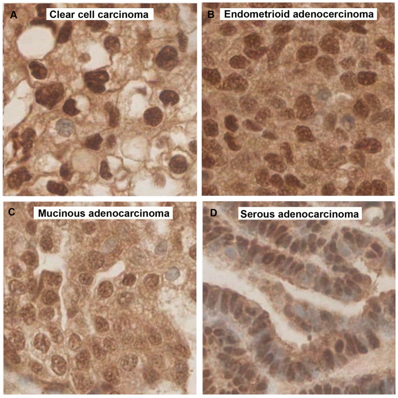
Mutations in the MCPH1 (Microcephalin) and ASPM (abnormal spindle-like microcephaly associated) genes cause primary microcephaly. Both are centrosomal associated proteins involved in mitosis. Microcephalin plays an important role in DNA damage response and ASPM is required for correct division of proliferative neuro-epithelial cells of the developing brain. Reduced MCPH1 mRNA expression and ASPM mRNA over-expression have been implicated in the development of human carcinomas. Epithelial ovarian cancer (EOC) is characterised by highly aneuploid tumours. Previously we have reported low Microcephalin and high ASPM protein levels and associations with clinico-pathological parameters in malignant cells from ascitic fluids. To confirm these previous findings on a larger scale Microcephalin and ASPM expression levels and localisations were evaluated by immunohistochemistry in two cohorts; a training set of 25 samples and a validation set of 322 EOC tissue samples. Results were correlated to the associated histopathological data. In normal ovarian tissues the Microcephalin nuclear staining pattern was consistently strong. In the cancer tissues, we identified low nuclear Microcephalin expression in high grade and advanced stage tumours (p<0.0001 and p = 0.0438 respectively). ASPM had moderate to high nuclear and low to moderate cytoplasmic expression in normal tissue. Cytoplasmic ASPM expression decreased with tumour grade and stage in the serous subtype of EOC (p = 0.023 and p = 0.011 respectively). Cytoplasmic ASPM increased with tumour stage in the endometrioid subtype (p = 0.023). Increasing tumour invasiveness (T3) and lymph node involvement (N1) also correlated with a decrease in cytoplasmic ASPM in EOC (p = 0.02 and p = 0.04 respectively). We have validated previous findings of deregulated expression of Microcephalin and ASPM in EOC by confirming associations for low nuclear Microcephalin levels and high cytoplasmic ASPM levels in a larger scale tumour tissue study. Microcephalin and ASPM may prove useful biomarkers in EOC.


Conflict of interest statement

Competing Interests: The authors have declared that no competing interests exist.

Figures

Figure 1
Figure 1. Immunohistochemical analysis of Microcephalin and ASPM in normal tissue samples.
Normal ovarian epithelium demonstrating strong Microcephalin expression in both the nuclei and cytoplasm (A & D) and strong nuclear and moderate cytoplasmic ASPM expression (G & J). Normal Fallopian tube demonstrating heterogeneous moderate nuclear and weak cytoplasmic Microcephalin staining (B & E) and ASPM staining demonstrating strong nuclear and moderate cytoplasmic expression (H & K). Normal Endometrium showing heterogeneous strong nuclear and cytoplasmic Microcephalin staining (C & F) and strong nuclear and weak cytoplasmic ASPM staining (I & L). All images are x15 magnification.
Figure 2
Figure 2. Immunohistochemical analysis of Microcephalin and ASPM expression in malignant samples.
Serous adenocarcinoma TMA cores showing nuclear and cytoplasmic Microcephalin (A and B) and ASPM expression (C and D) respectively. White arrow shows nuclear staining and black arrow shows cytoplasmic staining. A and B are 20x, C and D are 40x magnification.
Figure 3
Figure 3. Microcephalin expression in EOC validation set samples correlates with tumour grade.
Ai. Normal ovarian epithelial tissue with high Microcephalin expression. Aii-iv. Adenocarcinoma TMA cores showing strong Microcephalin expression in a low grade tumour (Aii), moderate Microcephalin in grade 2 (Aiii) and low levels of nuclear Microcephalin expression in a high grade tumour (Aiv). All images are 40x magnification. B. The correlation between nuclear Microcephalin expression decreases with increasing tumor grade (p<0.0001 using an ANOVA test).
Figure 4
Figure 4. Microcephalin expression in EOC validation set samples inversely correlates with tumour stage.
Ai. Normal ovarian epithelial tissue demonstrating high Microcephalin expression levels. Aii-4iv Adenocarcinoma TMA cores showing strong Microcephalin (Aii), moderate Microcephalin (Aiii) and low levels of nuclear Microcephalin expression (Aiv) in stage 1, 2 and 3 tumours respectively. All images are 40x magnification. B. Weak Microcephalin levels in the nucleus are associated with advanced tumour stage (p = 0.0438 using an ANOVA test).
Figure 5
Figure 5. Microcephalin expression in different EOC subtypes.
A. Clear cell carcinoma and B. Endometrioid adenocarcinoma both showing strong Microcephalin expression. C. Mucinous adenocarcinoma and D. Serous adenocarcinoma both presenting weak Microcephalin expression. All images are x40 magnification.
Figure 6
Figure 6. Variation of ASPM nuclear and cytoplasmic staining in validation set EOC tissue samples.
A and B. TMA core with low nuclear and cytoplasmic ASPM expression. C and D. TMA core with high nuclear and cytoplasmic ASPM expression. White arrow indicates nuclear stain, black arrow indicates cytoplasmic stain. A and C are 6.2x and B, D are 20x magnification respectively.
Figure 7
Figure 7. Cytoplasmic ASPM levels correlate with various clinic-pathological parameters in EOC.
A. Cytoplasmic ASPM levels decrease with grade in serous EOC (continuous data, p<0.0001). B. Cytoplasmic ASPM levels decrease with tumour grade in the serous subtype (discontinuous data, p = 0.0239). C. Cytoplasmic ASPM levels increase with disease stage in endometrioid EOC (discontinuous data, p = 0.0229). D. Cytoplasmic ASPM levels decrease with disease stage in the serous subtype (discontinuous data, p = 0.0017). E. Cytoplasmic ASPM levels decrease with tumour invasion (discontinuous data, p = 0.02). F. High cytoplasmic ASPM levels correlate with no lymph node involvement (p = 0.04). All data analysed using an ANOVA test.

References

    1. Ferlay J, Shin HR, Bray F, Forman D, Mathers C, et al... GLOBOCAN 2008 v1.2, Cancer Incidence and Mortality Worldwide.
    1. IARC Cancer base No. 10 [Internet] Lyon, France: International Agency for Research on Cancer, 2010. Available: http://globocan.iarc.fr.
    1. Auersperg N, Wong AS, Choi KC, Kang SK, Leung PC (2001) Ovarian surface epithelium: biology, endocrinology and pathology. Endocr Rev 22(2): 255–88. - PubMed
    1. Despierre E, Lambrechts D, Neven P, Amant F, Lambrechts S, et al. (2010) The molecular genetic basis of ovarian cancer and its roadmap towards a better treatment. Gynecol Oncol 117(2): 358–65. - PubMed
    1. Jackson AP, Eastwood H, Bell SM, Adu J, Toomes C, et al. (2002) Identification of microcephalin, a protein implicated in determining the size of the human brain. Am J Hum Genet 71(1): 136–42. - PMC - PubMed

Publication types

MeSH terms