. 2014 May 15;9(5):e97935.
doi: 10.1371/journal.pone.0097935. eCollection 2014.

Characterization of mannitol-2-dehydrogenase in Saccharina japonica: evidence for a new polyol-specific long-chain dehydrogenases/reductase

Affiliations

Characterization of mannitol-2-dehydrogenase in Saccharina japonica: evidence for a new polyol-specific long-chain dehydrogenases/reductase

Zhanru Shao et al. PLoS One. .

Abstract

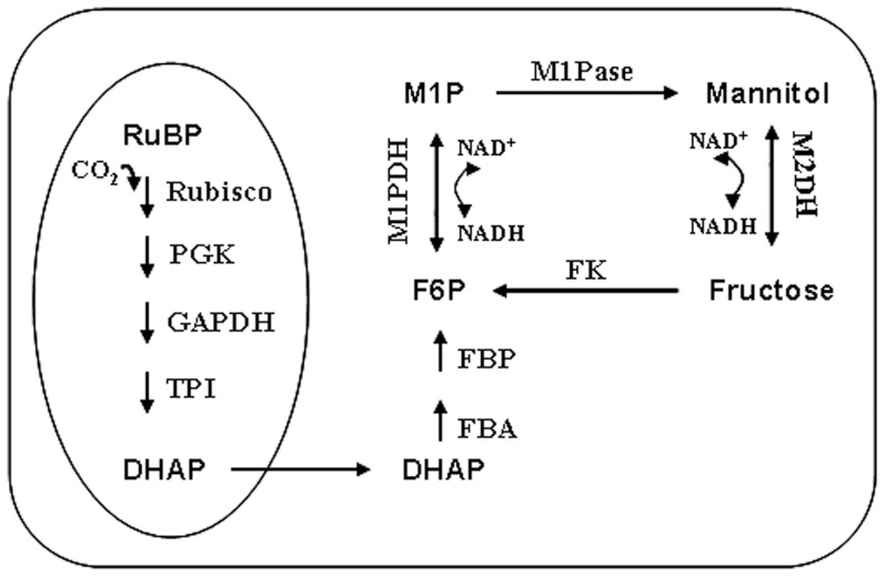
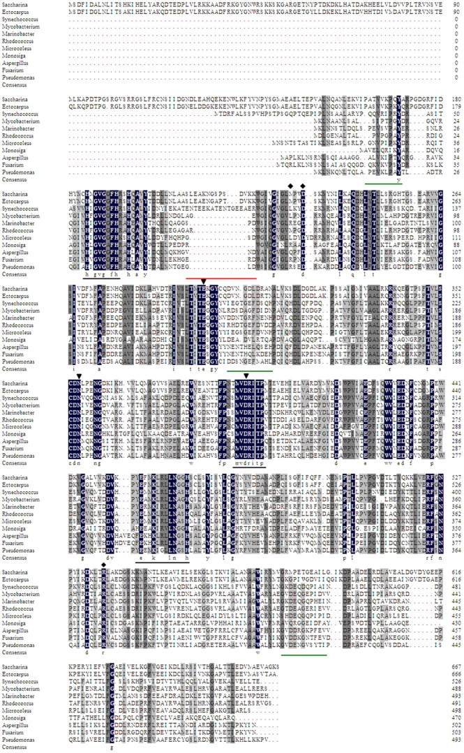
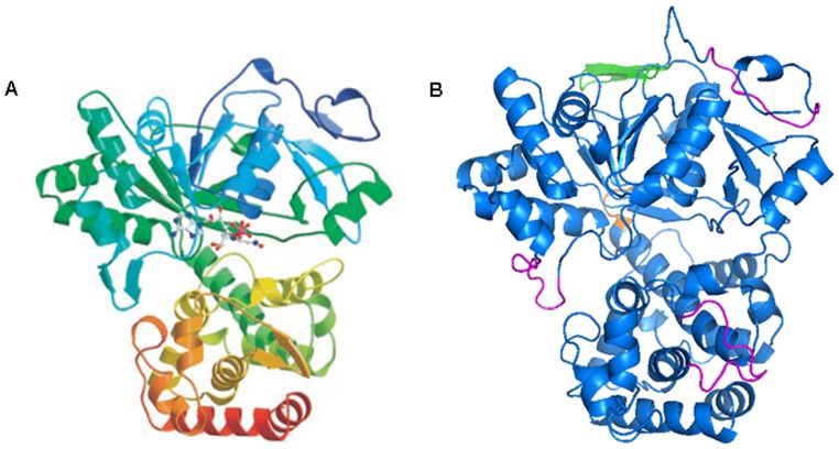
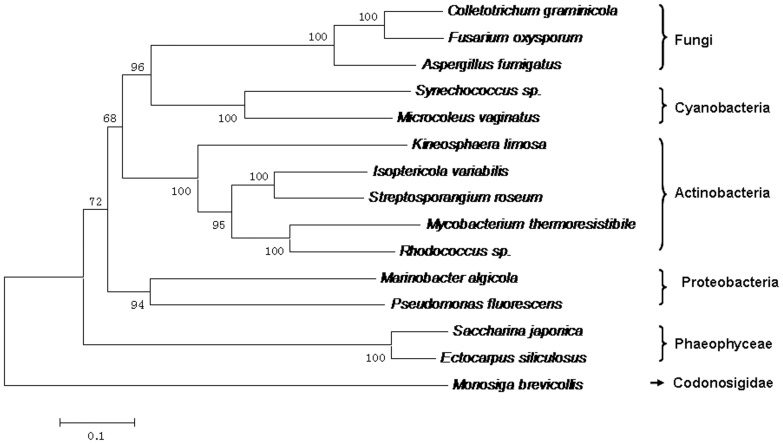
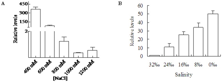
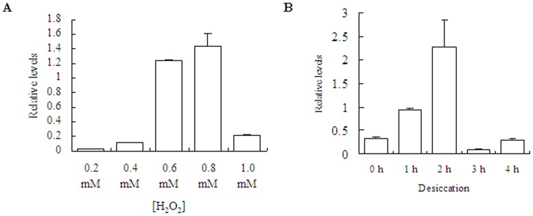
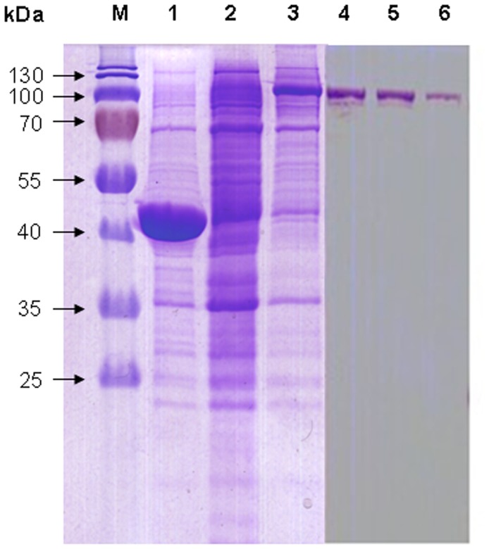
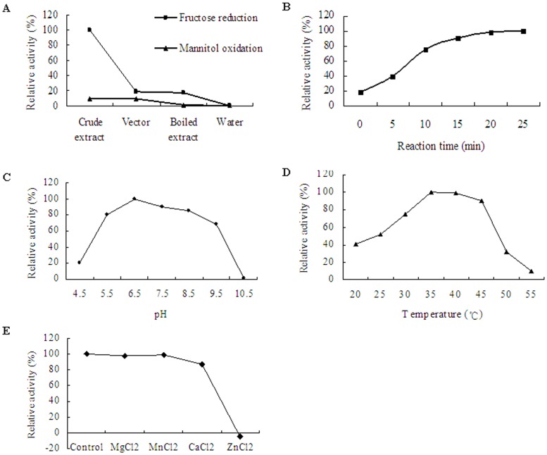
Mannitol plays a crucial role in brown algae, acting as carbon storage, organic osmolytes and antioxidant. Transcriptomic analysis of Saccharina japonica revealed that the relative genes involved in the mannitol cycle are existent. Full-length sequence of mannitol-2-dehydrogenase (M2DH) gene was obtained, with one open reading frame of 2,007 bp which encodes 668 amino acids. Cis-regulatory elements for response to methyl jasmonic acid, light and drought existed in the 5'-upstream region. Phylogenetic analysis indicated that SjM2DH has an ancient prokaryotic origin, and is probably acquired by horizontal gene transfer event. Multiple alignment and spatial structure prediction displayed a series of conserved functional residues, motifs and domains, which favored that SjM2DH belongs to the polyol-specific long-chain dehydrogenases/reductase (PSLDR) family. Expressional profiles of SjM2DH in the juvenile sporophytes showed that it was influenced by saline, oxidative and desiccative factors. SjM2DH was over-expressed in Escherichia coli, and the cell-free extracts with recombinant SjM2DH displayed high activity on D-fructose reduction reaction. The analysis on SjM2DH gene structure and biochemical parameters reached a consensus that activity of SjM2DH is NADH-dependent and metal ion-independent. The characterization of SjM2DH showed that M2DH is a new member of PSLDR family and play an important role in mannitol metabolism in S. japonica.


Conflict of interest statement

Competing Interests: The authors have declared that no competing interests exist.

Figures

Figure 1
Figure 1. Proposed pathway for photosynthetic carbon flow to the mannitol cycle in S. japonica.
The oval to the left represents the carbon fixation process which occurs in the chloroplast. RuBP, ribulose-1,5-bisphosphate; Rubisco, ribulose-1,5-bisphosphate carboxylase/oxygenase; PGK, phosphoglycerate kinase; GAPDH, glyceraldehyde-3-phosphate dehydrogenase; TPI, triose phosphate isomerase; DHAP, Dihydroxyacetone phosphate; FBA, fructose bisphosphate aldolase; FBP, fructose-1,6-bisphosphatase; F6P, fructose-6-phosphate; M1P, mannitol-1-phosphate; M1PDH, mannitol-1-phosphate dehydrogenase; M1Pase, mannitol-1-phosphatase; M2DH, mannitol-2-dehydrogenase; FK, fructokinase.
Figure 2
Figure 2. Multiple sequence alignment of MDHs from typical species of Phaeophyceae, Cyanobacteria, Actinobacteria, Proteobacteria, Ascomycota and Codonosigidae.
Identical residues among all MDHs were shown in black boxes. The representative conserved regions among PSLDRs were underlined in black. The deletions of β-sheets in SjM2DH were underlined in green while the extra anti-parallel β-sheet was underlined in red. ▾, residues for substrate-binding; ⧫, residues for NADH-binding. The accession numbers were listed in File S2.
Figure 3
Figure 3. Structural comparison of the crystal structure of PfM2DH and putative SjM2DH.
A, global tertiary structure of PfM2DH (Kavanagh et al., 2002). B, stereo-ribbon representation of SjM2DH in two domains. The connection of N- and C-terminal domains (VKDV) was indicated in orange; three deletions of β-sheets were shown in magenta; the insertion of an anti-parallel β-sheet was presented in green.
Figure 4
Figure 4. Phylogenetic tree constructed based on alignment of 15 M2DH amino acid sequences.
The tree was obtained by the neighbor-joining algorithm using the MEGA 5.2 program. Bootstrap values calculated from 1,000 replicates were given. The scale bar corresponded to 0.1 estimated amino-acid substitutions per site.
Figure 5
Figure 5. Influence of salinities and NaCl concentrations on SjM2DH expression levels in juvenile sporophytes.
A, expression levels of SjM2DH under various NaCl concentrations. B, expression levels of SjM2DH under various salinities. All the data are the mean values of three independent experiments.
Figure 6
Figure 6. Influence of H2O2 concentrations and desiccation on SjM2DH expression levels in juvenile sporophytes.
A, expression levels of SjM2DH under various H2O2 concentrations. B, expression levels of SjM2DH under different duration of desiccation stress. All the data are the mean values of three independent experiments.
Figure 7
Figure 7. SDS-PAGE (12%) analysis of induction and purification of recombinant SjM2DH.
M, protein marker; lanes 1 and 2, negative controls of untransformed vector and recombinant E. coli induced with 0 mM IPTG, respectively; lane 3, recombinant E. coli with 0.3 mM IPTG induction for 1 h; lanes 4–6, eluted fractions with presence of recombinant SjM2DH.
Figure 8
Figure 8. Enzymatic characteristics of recombinant SjM2DH in cell-free bacterial extracts.
A, relative activity in fructose reduction and mannitol oxidation directions. B, time-course evaluation (0–25 min) of reductive activity on fructose. C, influence of different pH (4.5–10.5) on the activity of SjM2DH. D, influence of temperature (20–55°C) on SjM2DH activity. E, influence of metal ion on the activity of SjM2DH.

References

    1. Wisselink HW, Weusthuis RA, Eggink G, Hugenholtz J, Grobben GJ (2002) Mannitol production by lactic acid bacteria: a review. International Dairy Journal 12: 151–161.
    1. Hörer S, Stoop J, Mooibroek H, Boumann U, Sassoon J (2001) The crystallographic structure of the mannitol 2-dehydrogenase NADP+ binary complex from Agaricus bisporus . The Journal of Biological Chemistry 276: 27555–27561. - PubMed
    1. Lee JK, Koo BS, Kim SY, Hyun HH (2003) Purification and characterization of a novel mannitol dehydrogenase from a newly isolated strain of Candida magnoliae . Applied and Environmental Microbiology 69 8: 4438–4447. - PMC - PubMed
    1. Stoop JMH, Williamson JD, Pharr DM (1996) Mannitol metabolism in plants: a method for coping with stress. Trends in Plant Science 1: 139–144.
    1. Yamaguchi T, Ikawa T, Nisizawa K (1969) Pathway of mannitol formation during photosynthesis in brown algae. Plant and Cell Physiology 10: 425–440.

Publication types