. 2014 Jul 11;289(28):19351-63.
doi: 10.1074/jbc.M114.558882. Epub 2014 May 15.

Tyrosine 308 is necessary for ligand-directed Gs protein-biased signaling of β2-adrenoceptor

Affiliations

Tyrosine 308 is necessary for ligand-directed Gs protein-biased signaling of β2-adrenoceptor

Anthony Yiu-Ho Woo et al. J Biol Chem. .

Abstract

Interaction of a given G protein-coupled receptor to multiple different G proteins is a widespread phenomenon. For instance, β2-adrenoceptor (β2-AR) couples dually to Gs and Gi proteins. Previous studies have shown that cAMP-dependent protein kinase (PKA)-mediated phosphorylation of β2-AR causes a switch in receptor coupling from Gs to Gi. More recent studies have demonstrated that phosphorylation of β2-AR by G protein-coupled receptor kinases, particularly GRK2, markedly enhances the Gi coupling. We have previously shown that although most β2-AR agonists cause both Gs and Gi activation, (R,R')-fenoterol preferentially activates β2-AR-Gs signaling. However, the structural basis for this functional selectivity remains elusive. Here, using docking simulation and site-directed mutagenesis, we defined Tyr-308 as the key amino acid residue on β2-AR essential for Gs-biased signaling. Following stimulation with a β2-AR-Gs-biased agonist (R,R')-4'-aminofenoterol, the Gi disruptor pertussis toxin produced no effects on the receptor-mediated ERK phosphorylation in HEK293 cells nor on the contractile response in cardiomyocytes expressing the wild-type β2-AR. Interestingly, Y308F substitution on β2-AR enabled (R,R')-4'-aminofenoterol to activate Gi and to produce these responses in a pertussis toxin-sensitive manner without altering β2-AR phosphorylation by PKA or G protein-coupled receptor kinases. These results indicate that, in addition to the phosphorylation status, the intrinsic structural feature of β2-AR plays a crucial role in the receptor coupling selectivity to G proteins. We conclude that specific interactions between the ligand and the Tyr-308 residue of β2-AR stabilize receptor conformations favoring the receptor-Gs protein coupling and subsequently result in Gs-biased agonism.

Keywords: Adrenergic Receptor; Cardiomyocyte Contraction; Cardiovascular; Functional Selectivity; G Protein-coupled Receptor (GPCR); Molecular Docking; Molecular Pharmacology; Signal Transduction; Site-directed Mutagenesis.


Figures

FIGURE 1.
FIGURE 1.
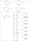
Structures of Fen and its derivatives used in the study. The following terms are used: fenoterol (Fen), 4′-methoxyfenoterol (methoxyFen); 4′-aminofenoterol (aminoFen); phenylfenoterol (PhFen); 1-naphthylfenoterol (1-NapFen); ethylfenoterol (EtFen); 2-naphthylfenoterol (2-NapFen); and 4′-methoxy-1-naphthylfenoterol (MNFen).
FIGURE 2.
FIGURE 2.
Substitution on the aminoalkyl portion of (R,R′)-Fen determines the PTX sensitivity of the agonist-stimulated contractile response in rat cardiomyocytes. Concentration-response profiles of cardiomyocyte contractility in cells subjected to (R,R′)-PhFen (A), (R,R′)-aminoFen (B), (R,R′)-1-NapFen (C), and (R,R′)-MNFen (D) with (▴) and without (○) PTX treatment (0.75 μg/ml at 37 °C for >3 h). Contractile response to the agonist is expressed as a percentage of the basal contractility (mean ± S.E., n = 9–11 cells from 5 to 9 hearts for each data point).
FIGURE 3.
FIGURE 3.
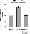
(R,R′)-AminoFen exhibits β2-AR subtype selectivity in cardiomyocyte contractile response. Single ventricular myocytes from rats were set to pace under perfusion. The contraction amplitude of a cell in response to (R,R′)-aminoFen (0.1 μm) followed by ICI-118,551 (ICI, 0.1 μm) was monitored. Steady-state contractility was recorded. Contractile response is expressed as a percentage of the basal contractility. Data are means ± S.E., n = 4 cells from four hearts. ***, p < 0.001 (by paired t test).
FIGURE 4.
FIGURE 4.
Addition of forskolin reconstitutes functional coupling of β2-AR to Gi protein in cultured β2-AR knock-out mouse cardiomyocytes induced with human β2-AR. Cardiomyocytes from β2-AR knock-out mice were infected with adeno-GFP (white bar) or adeno-β2-AR (black bars) and cultured for 24 h in the presence or absence of forskolin (1 μm) and/or PTX (0.75 μg/ml) as indicated. Cells were transferred to a perfusion chamber, electrically paced, and subjected to stimulation with zinterol (0.2 μm, a concentration without an inotropic effect in freshly isolated cardiomyocytes from WT mice, see Fig. 1A in Ref. 26). Steady-state contractility was measured. Data (mean ± S.E., n = 10–15 cells from 5 to 9 hearts for each data point) are expressed as percentages of the basal contractility. *, p < 0.05. Zinterol (0.2 μm) did not increase contractility in cells infected with adeno-GFP demonstrating no β1-AR stimulatory effect at this concentration. In cells infected with adeno-β2-AR and cultured in the absence of forskolin, the inotropic response produced by zinterol stimulation was the result of a pure β2-AR-Gs-mediated effect because β2-AR and Gi proteins were functionally uncoupled. In cells infected with adeno-β2-AR in the presence of forskolin, the coupling of β2-AR to Gi protein was reestablished. Therefore, the cardiomyocytes were unresponsive to zinterol as if they were freshly isolated WT β2-AR+ cells when β2-AR-Gi coupling was intact. In cells infected with adeno-β2-AR in the presence of forskolin and PTX, the coupling of β2-AR to Gi protein still occurred, but Gi had lost its function and could no longer negatively regulate β2-AR-Gs activation by zinterol.
FIGURE 5.
FIGURE 5.
PTX increases the inotropic effect of (R,R′)-aminoFen in cardiomyocytes expressing β2-AR Y308F mutant but not in cardiomyocytes expressing WT human β2-AR. A, (R,R′)-aminoFen-stimulated contractile responses in β2-AR knock-out mouse cardiomyocyte adenoviral gene transfer GFP. Cardiomyocytes from β2-AR knock-out mice were infected with adeno-GFP and cultured for 24 h. Cells were paced under perfusion and subjected to (R,R′)-aminoFen (100, 500, or 1000 nm). Steady-state contractility before and after agonist stimulation was measured. B, (R,R′)-aminoFen-stimulated contractile responses in β2-AR knock-out mouse cardiomyocytes adenoviral gene transfer WT β2-AR in the presence or absence of PTX treatment. Cardiomyocytes from β2-AR knock-out mice were infected with adeno-β2-AR and cultured with or without PTX (0.75 μg/ml) for 24 h. Cells were paced under perfusion and subjected to (R,R′)-aminoFen (10, 100, or 500 nm). C, (R,R′)-aminoFen-stimulated contractile responses in β2-AR knock-out mouse cardiomyocyte adenoviral gene transfer β2-AR Y308F in the presence or absence of PTX treatment. Contractile responses are expressed as percentages of the basal contractility (mean ± S.E., n = 9–14 cells from 4 to 8 hearts for each data point). Concentration dependence of the (R,R′)-aminoFen-stimulated responses was verified (by two-way analysis of variance, p < 0.0001) for all datasets. *, p < 0.05; ***, p < 0.001 versus corresponding −PTX group.
FIGURE 6.
FIGURE 6.
Y308F substitution on β2-AR increases the PTX sensitivity of (R,R′)-aminoFen-induced ERK phosphorylation in HEK stable cell lines. Confluent cultures of HEK-β2-AR cells and HEK-β2-AR Y308F cells were deprived of serum overnight. Treatment with PTX (0.3 μg/ml, +) or vehicle (−) was implemented during serum starvation. Cells were then stimulated with ISO (1 μm) or (R,R′)-aminoFen (10−9 to 10−6 m) for 5 min at 37 °C as indicated. ERK phosphorylation was determined by immunoblotting. A, immunoblots of p-ERK and total ERK (as protein loading control) in response to agonist stimulation in HEK-β2-AR cells, and B, averaged data. C, immunoblots of p-ERK and total ERK in response to agonist stimulation in HEK-β2-AR Y308F cells, and D, averaged data. Data are presented as fold increase over −PTX control (means ± S.E. in 3–4 independent experiments). *, p < 0.05; **, p < 0.01; ***, p < 0.001 versus vehicle controls; #, p < 0.05 versus −PTX group.
FIGURE 7.
FIGURE 7.
Y308F substitution on β2-AR increases the PTX sensitivity of (R,R′)-aminoFen-stimulated cAMP production in HEK stable cell lines. HEK-β2-AR cells and HEK-β2-AR Y308F cells were cultured in 12-well plates in parallel, and subsets of the cells were treated with PTX (0.3 μg/ml) or vehicle overnight. Agonist stimulation was allowed to proceed for 10 min at 25 °C in the presence of 3-isobutyl-1-methylxanthine (1 mm). Cellular cAMP contents were determined by enzyme immunoassay. HEK-β2-AR cells were subjected to (R,R′)-aminoFen (10−11 to 10−6 m) (A), and HEK-β2-AR Y308F cells were subjected to (R,R′)-aminoFen (B), with (▴) and without (○) PTX treatment. Data (means ± S.E. in three independent experiments performed in triplicate) are expressed as percentages of the Emax response of the β2-AR WT −PTX group. Curve-fitting analysis of the concentration-response curves were conducted using Prism. **, p < 0.01 versus −PTX group.
FIGURE 8.
FIGURE 8.
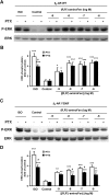
(R,R′)-AminoFen induces phosphorylation of β2-AR and β2-AR Y308F mutant at the GRK and the PKA sites in HEK stable cell lines. Confluent cultures of HEK-β2-AR cells and HEK-β2-AR Y308F cells were incubated in serum-free medium for 3 h and then stimulated with vehicle control (−), ISO (1 μm), or (R,R′)-aminoFen (R, 1 μm; R100 for WT, 0.2 μm; R100 for Y308F, 1 μm; R1000 for WT, 2 μm; R1000 for Y308F, 10 μm) for 5 min at 37 °C. Phosphorylated β2-AR was detected by phosphosite-specific antibodies against Ser(P)-262 for PKA sites and Ser(P)-355,356 for GRK sites. Total β2-AR was detected after stripping and reprobing the membrane with the β2-AR-CT antibody. A, immunoblots of Ser(P)-262-β2-AR and total β2-AR in response to agonist stimulation, and B, averaged data (normalized to total β2-AR). C, immunoblots of Ser(P)-355,356-β2-AR and total β2-AR in response to agonist stimulation, and D, averaged data. Data are expressed as fold increase over control (means ± S.E. in at least three independent experiments). *, p < 0.05; **, p < 0.01; ***, p < 0.001 versus vehicle controls (two-way analysis of variance with post hoc t test). No significant differences were found for all within-group comparisons between WT and Y308F, p > 0.05.
FIGURE 9.
FIGURE 9.
Binding poses of (R,R′)-aminoFen and (R,R′)-MNFen docked to the β2-AR models. A, docking of (R,R′)-aminoFen to the carazolol-bound β2-AR model (PDB entry 2RH1), and B, to the β2-AR Y308F mutant. The 4′-amino group of (R,R′)-aminoFen forms a HB with the Tyr-308 residue of β2-AR (green arrow), and the interaction is lost in the β2-AR Y308F mutant. C, docking of (R,R′)-aminoFen to a BI-167107 and Gs protein-bound conformation of β2-AR (PDB entry 3SN6). The location of the docked molecule highly resembles the orientation of co-crystallized agonist, BI-167107, as both molecules share significant structural similarities. The agonists form a network of analogous HB interactions with the receptor residues Ser-203, Ser-207, Asn-312, and Asp-113. An additional interaction can be observed between the 4′-amino moiety of the ligand and Lys-305 residue. D, docking of (R,R′)-MNFen to a carazolol-bound conformation of β2-AR (PDB entry 2RH1). Aromatic residues in the ligand binding pocket able to form π-π interactions with the naphthyl moiety of the ligand are shown. Ligand molecule is rendered in atom-type color-coded stick mode, and five essential TM helices of target β2-AR model are shown and colored as follows: TM3, magenta; TM4, green; TM5, red; TM6, yellow; and TM7, blue.

Similar articles

Cited by

References

    1. Violin J. D., Lefkowitz R. J. (2007) β-Arrestin-biased ligands at seven-transmembrane receptors. Trends Pharmacol. Sci. 28, 416–422 - PubMed
    1. Kenakin T. (2007) Collateral efficacy in drug discovery: taking advantage of the good (allosteric) nature of 7TM receptors. Trends Pharmacol. Sci. 28, 407–415 - PubMed
    1. Mailman R. B. (2007) GPCR functional selectivity has therapeutic impact. Trends Pharmacol. Sci. 28, 390–396 - PMC - PubMed
    1. Urban J. D., Clarke W. P., Zastrow M. V., Nichols D. E., Kobilka B., Weinstein H. (2007) Functional selectivity and classical concepts of quantitative pharmacology. Pharmacology 320, 1–13 - PubMed
    1. Neubig R. R. (2007) Missing links: mechanisms of protean agonism. Mol. Pharmacol. 71, 1200–1202 - PubMed

Publication types

MeSH terms

Substances