. 2014 May 15;8(5):e2856.
doi: 10.1371/journal.pntd.0002856. eCollection 2014 May.

Hemoglobin uptake by Paracoccidioides spp. is receptor-mediated

Affiliations

Hemoglobin uptake by Paracoccidioides spp. is receptor-mediated

Elisa Flávia Luiz Cardoso Bailão et al. PLoS Negl Trop Dis. .

Abstract

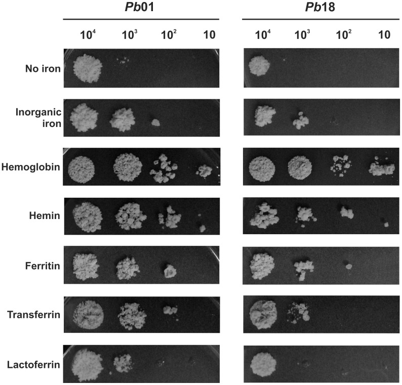
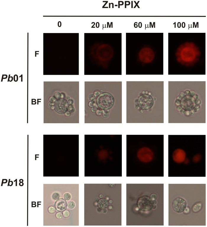
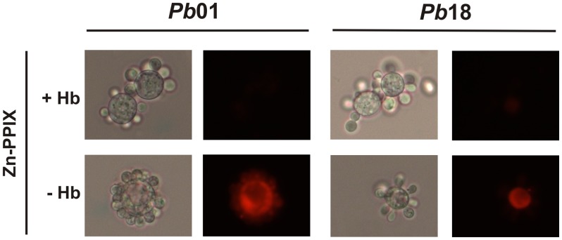
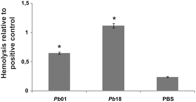
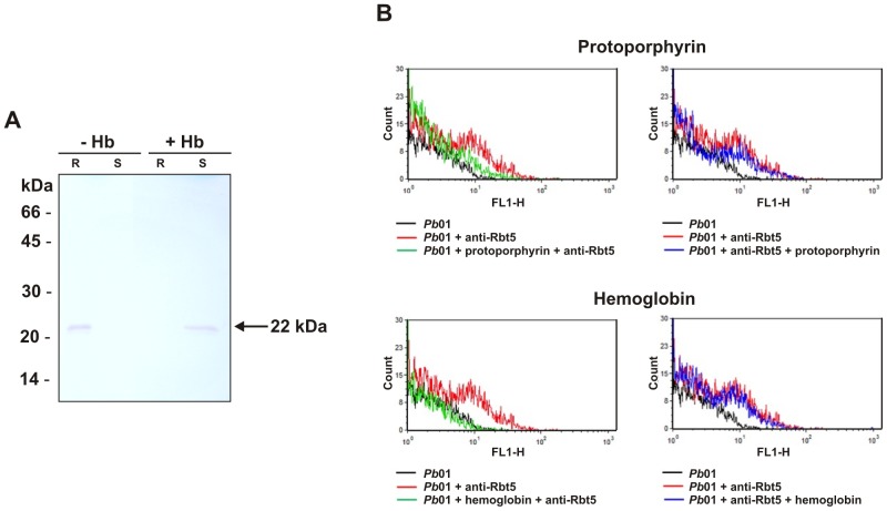
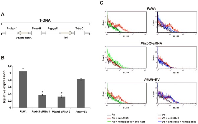
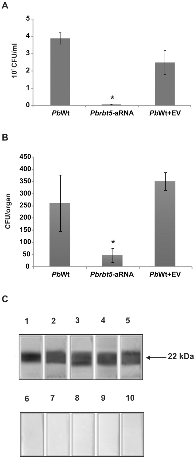
Iron is essential for the proliferation of fungal pathogens during infection. The availability of iron is limited due to its association with host proteins. Fungal pathogens have evolved different mechanisms to acquire iron from host; however, little is known regarding how Paracoccidioides species incorporate and metabolize this ion. In this work, host iron sources that are used by Paracoccidioides spp. were investigated. Robust fungal growth in the presence of the iron-containing molecules hemin and hemoglobin was observed. Paracoccidioides spp. present hemolytic activity and have the ability to internalize a protoporphyrin ring. Using real-time PCR and nanoUPLC-MSE proteomic approaches, fungal growth in the presence of hemoglobin was shown to result in the positive regulation of transcripts that encode putative hemoglobin receptors, in addition to the induction of proteins that are required for amino acid metabolism and vacuolar protein degradation. In fact, one hemoglobin receptor ortholog, Rbt5, was identified as a surface GPI-anchored protein that recognized hemin, protoporphyrin and hemoglobin in vitro. Antisense RNA technology and Agrobacterium tumefaciens-mediated transformation were used to generate mitotically stable Pbrbt5 mutants. The knockdown strain had a lower survival inside macrophages and in mouse spleen when compared with the parental strain, which suggested that Rbt5 could act as a virulence factor. In summary, our data indicate that Paracoccidioides spp. can use hemoglobin as an iron source most likely through receptor-mediated pathways that might be relevant for pathogenic mechanisms.


Conflict of interest statement

The authors have declared that no competing interests exist.

Figures

Figure 1
Figure 1. Effect of different iron sources on the growth of Paracoccidioides yeast cells.
Pb01 and Pb18 cell cultures were collected after 36 h of iron scarcity, washed and ten-fold serial dilutions of cell suspensions (104 to 10 cells) were spotted on MMcM medium plates, which were supplemented with 50 µM BPS, an iron chelator. As indicated, different iron sources were added or not (no iron condition): 30 µM inorganic iron, 30 µM hemoglobin, 120 µM hemin, 30 µg/µl ferritin, 30 µM transferrin or 3 µM lactoferrin.
Figure 2
Figure 2. Paracoccidioides can internalize protoporphyrin rings.
Iron deprived Pb01 and Pb18 yeast cells were incubated in MMcM medium supplemented or not (0) with different zinc protoporphyrin IX (Zn-PPIX) concentrations (20–100 µM) for 2 h. After this period, the cells were washed twice, and observed by bright field microscopy (BF) and by live fluorescence microscopy (F).
Figure 3
Figure 3. Hemoglobin can block the Zn-PPIX internalization by Paracoccidioides.
Iron deprived Pb01 and Pb18 yeast cells were pre-incubated (+) or not (−) with hemoglobin (Hb) for 1 h. After, the cells were incubated in MMcM medium supplemented with 60 µM zinc protoporphyrin IX (Zn-PPIX) for 2 h. After this period, the cells were washed twice and observed by bright field microscopy (left panels for each strain) and by live fluorescence microscopy (right panels for each strain).
Figure 4
Figure 4. Hemolysis of sheep erythrocytes in the presence of Paracoccidioides yeast cells.
Pb01 and Pb18 107 yeast cell suspensions were incubated with 108 sheep erythrocytes for 2 h at 36°C in 5% CO2. As a negative or positive control, respectively, erythrocytes were incubated with phosphate buffered saline solution (PBS) or sterile water. The optical densities of the supernatants were determined with an ELISA plate reader at 405 nm. The experiment was performed in triplicate, and the average optical density of each condition was used to calculate the relative hemolysis of the experimental conditions or the negative control against the positive control. The data are plotted as the mean ± standard deviation. *: statistically significant difference in comparison with PBS values according to Student's t-test.
Figure 5
Figure 5. Expression of genes that are putatively related to hemoglobin uptake.
Pb01 yeast cells were recovered from MMcM medium, which was supplemented or not (no iron addition condition) with different iron sources (10 µM and 100 µM inorganic iron and 10 µM hemoglobin) for 30, 60 and 120 min. After RNA extraction and cDNA synthesis, levels of Pb01 rbt5, wap1 and csa2 transcripts were quantified by qRT-PCR. The expression values were calculated using alpha tubulin as the endogenous control. The values that were plotted on the bar graph were normalized against the expression data that were obtained from the no iron addition condition (fold change). The data are expressed as the mean ± SD of the triplicates. *statistically significant data as determined by Student's t-test (p<0.05).
Figure 6
Figure 6. Paracoccidioides Rbt5 is a GPI-anchored protein localized in the yeast cell wall.
A. Cell wall fraction of Pb01 yeast cells was obtained and analyzed by Western blot using polyclonal antibodies raised against the recombinant protein Rbt5. Proteins that were obtained from the cell wall (lane 1) were extracted by HF-pyridine digestion and analyzed (lane 2). Molecular weight markers are indicated at the right side of the panel. B. Immunoelectron microscopic detection of Rbt5 in Pb01 yeast cells by post embedding methods. (1) Negative control exposed to the rabbit preimmune serum. (2 and 3) Gold particles are observed at the fungus cell wall (arrow) and in the cytoplasm (double arrowheads). Bars: 1 µm (1 and 2) and 0.5 µm (3). v: vacuoles. m: mitochondria. w: cell wall.
Figure 7
Figure 7. Paracoccidioides Rbt5 binds heme-containing molecules.
A. Recombinant protein Rbt5 was pre-incubated with (+) or without (−) hemoglobin (Hb) for 1 h. Subsequently, the samples were incubated with hemin-agarose resin for 1 h. After, the samples were centrifuged, the supernatants (S) were collected and the resin (R) was washed twice. After adding the buffer, the samples were boiled for 5 min and submitted to SDS-PAGE and Western blot analysis. A single 22 kDa immunoreactive species, which corresponds to the Rbt5 recombinant protein, was detected bound to the resin in the absence of hemoglobin or in the supernatant in the presence of hemoglobin. B. Upper and lower panels represent systems where protoporphyrin or hemoglobin Rbt5 recognition was assessed, respectively. The sequential steps of incubation in each system are indicated on the bottom of each panel. Pb01 denotes the background fluorescence of fungal cells alone; Pb01 + anti-Rbt5 represents systems where fungal cells were sequentially incubated with primary and secondary antibodies; Pb01 + protoporphyrin/hemoglobin + anti-Rbt5 is representative of systems that included the blocking of yeast cells with heme-containing molecules before exposure to antibodies; and Pb01 + anti-Rbt5 + protoporphyrin/hemoglobin represents systems that included the incubation of yeast cells with heme-containing molecules after exposure to antibodies.
Figure 8
Figure 8. Paracoccidioides Rbt5 knock down via an antisense-RNA (aRNA) strategy.
A. Schematic representation of the T-DNA cassette that was used in this work to perform the Agrobacterium tumefaciens-mediated transformation (ATMT) of Pb339 (PbWt). Pbrbt5-aRNA was cloned in the pUR5750 binary vector under the control of the Histoplasma capsulatum cbp-1 gene promoter region (P-cbp-1) and the Aspergillus fumigatus cat-B gene termination region (T-cat-B). The selection marker that was used in this work was the Escherichia coli hygromycin-resistance gene hph. In the cassette, this gene is flanked by the glyceraldehyde-3-phosphate dehydrogenase promoter region (P-gapdh) and by the trpC termination region (T-trpC) from Aspergillus nidulans. B. After the selection of mitotic stable isolates, a qRT-PCR was performed to analyze the silencing level of the gene in isolates that were transformed with Pbrbt5-aRNA. As controls, rbt5 transcript level from PbWt and PbWt transformed with the empty vector (PbWt+EV) were also quantified. Alpha tubulin was used as the endogenous control. The data are represented as the means ± SD from triplicate determinations. *: statistically significant data as determined by Student's t-test (p<0.05) in comparison with the data that were obtained from PbWt+EV strain. C. Effect of Pbrbt5 deletion on the interaction of Paracoccidioides with heme-containing molecules. Hemoglobin prevents PbWt and PbWt+EV cells to be recognized by the anti-Rbt5 antibodies. However, Pbrbt5-aRNA cells are poorly recognized by the antibody that was raised against Rbt5, which is a process that was not affected by the previous or subsequent exposure of yeast cells to hemoglobin.
Figure 9
Figure 9. Paracoccidioides Rbt5 shows virulent and antigenic properties.
A. To test the ability to infect macrophages, PbWt, Pbrbt5-aRNA or PbWt+EV strains were co-cultivated with macrophages for 24 h. After this period, infected macrophages were lysed, and lysates were plated on BHI medium to recover the fungi. The data are presented as a bar graph of the means ± SEM from triplicates. *: statistically significant data as determined by Student's t-test (p<0.05) in comparison with the data that were obtained from the PbWt+EV strain. B. A murine model of infection was also used. Mice were infected intraperitoneally with PbWt, Pbrbt5-aRNA or PbWt+EV strains. After 2 weeks of infection, mice were sacrificed, the spleens were removed and samples of the homogenate were plated on BHI medium. After 15 days, the CFUs were counted to determine the fungal burden for each strain. The data are presented as a bar graph of the means ± SEM from quadruplicates. *: statistically significant data as determined by Student's t-test (p<0.05) in comparison with the data that were obtained from the PbWt+EV strain. C. Reaction of the recombinant Pb01 Rbt5 with sera of five PCM patients (lanes 1–5) or with control sera (lanes 6–10). After reacting with the anti-human IgG peroxidase coupled antibody, the reaction was developed using hydrogen peroxide and diaminobenzidine.

References

    1. Schrettl M, Haas H (2011) Iron homeostasis—Achilles' heel of Aspergillus fumigatus? Curr Opin Microbiol 14: 400–405. - PMC - PubMed
    1. Kaplan CD, Kaplan J (2009) Iron acquisition and transcriptional regulation. Chem Rev 109: 4536–4552. - PubMed
    1. Nevitt T (2011) War-Fe-re: iron at the core of fungal virulence and host immunity. Biometals 24: 547–558. - PubMed
    1. Ramakrishna G, Rooke TW, Cooper LT (2003) Iron and peripheral arterial disease: revisiting the iron hypothesis in a different light. Vasc Med 8: 203–210. - PubMed
    1. Hentze MW, Muckenthaler MU, Galy B, Camaschella C (2010) Two to tango: regulation of Mammalian iron metabolism. Cell 142: 24–38. - PubMed

Publication types

LinkOut - more resources