Development of external surfaces of human cerebellar lobes in the fetal period
- PMID: 24831768
- PMCID: PMC4155164
- DOI: 10.1007/s12311-014-0566-3
Development of external surfaces of human cerebellar lobes in the fetal period
Abstract
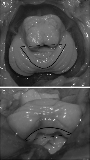
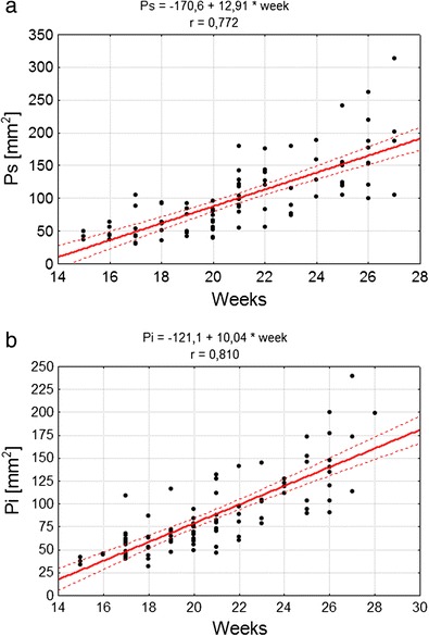
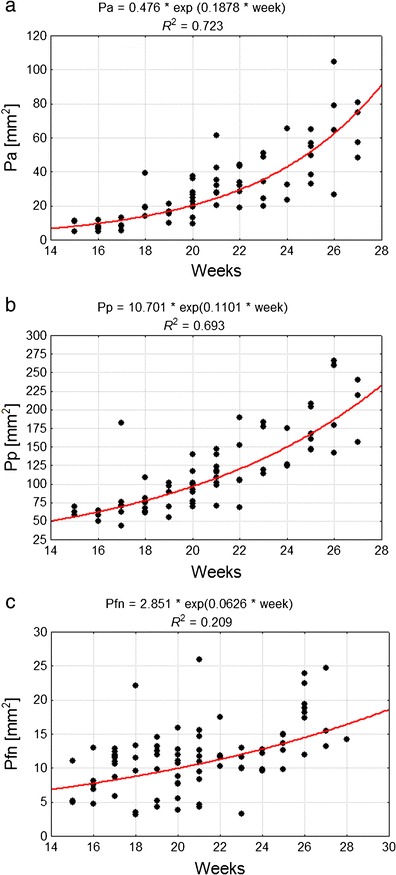
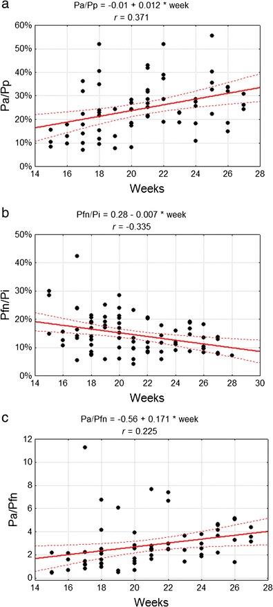
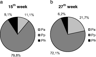
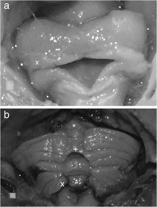
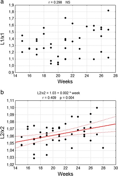
In the fetal period, development of cerebellar lobes may proceed dissimilarly due to possible differentiated origins of the cells and diversified times of their migration to certain cerebellum regions. This can cause various growth trajectories for the external surfaces of cerebellar lobes. The goal of the study was to describe the development of the external surface of cerebellum lobes and fissures delineating them in the fetal period. The material consisted of 101 fetuses (48 males and 53 females)-crown rump length 89-229 mm corresponding to 15-28 weeks of fetal life. The methods were based on anthropometric measurements and preparation techniques combined with elicited image computer analysis. At the largest values of the cerebellum posterior lobe surface, the most dynamic growth rate was observed in the case of the anterior lobe. Among the cerebellar lobes, proportional change was observed as well as a gradual increase in anterior lobe surface area and a simultaneous decrease in the surface area of the flocculonodular lobe part of the cerebellum total external surface. This paper presents the different growth trajectories of cerebellar lobes and demonstrates the importance of the primary fissure as a delineating mark for two regions with different dynamics of development.
Figures


Similar articles
-
3D morphometric analysis of human fetal cerebellar development.Cerebellum. 2012 Sep;11(3):761-70. doi: 10.1007/s12311-011-0338-2. Cerebellum. 2012. PMID: 22198870 Free PMC article.
-
Mathematical models of human cerebellar development in the fetal period.J Anat. 2018 Apr;232(4):596-603. doi: 10.1111/joa.12767. Epub 2018 Jan 8. J Anat. 2018. PMID: 29315634 Free PMC article.
-
MR volume of the fetal cerebellum in relation to growth.J Magn Reson Imaging. 2008 Apr;27(4):840-5. doi: 10.1002/jmri.21290. J Magn Reson Imaging. 2008. PMID: 18302203
-
Magnetic resonance imaging of fetal cerebellar development.Cerebellum. 2006;5(3):199-205. doi: 10.1080/14734220600589210. Cerebellum. 2006. PMID: 16997751 Review.
-
MRI of fetal and neonatal cerebellar development.Semin Fetal Neonatal Med. 2005 Oct;10(5):411-20. doi: 10.1016/j.siny.2005.05.004. Semin Fetal Neonatal Med. 2005. PMID: 15985392 Review.
Cited by
-
Kola nut from Cola nitida vent. Schott administered to pregnant rats induces histological alterations in pups' cerebellum.PLoS One. 2021 Mar 8;16(3):e0247573. doi: 10.1371/journal.pone.0247573. eCollection 2021. PLoS One. 2021. PMID: 33684143 Free PMC article.
-
A mutation in the THG1L gene in a family with cerebellar ataxia and developmental delay.Neurogenetics. 2016 Oct;17(4):219-225. doi: 10.1007/s10048-016-0487-z. Epub 2016 Jun 15. Neurogenetics. 2016. PMID: 27307223
-
Morphometric development of the human fetal cerebellum during the early second trimester.Neuroimage. 2020 Feb 15;207:116372. doi: 10.1016/j.neuroimage.2019.116372. Epub 2019 Nov 18. Neuroimage. 2020. PMID: 31751665 Free PMC article.
-
Cellular commitment in the developing cerebellum.Front Cell Neurosci. 2015 Jan 12;8:450. doi: 10.3389/fncel.2014.00450. eCollection 2014. Front Cell Neurosci. 2015. PMID: 25628535 Free PMC article. Review.
-
Hindbrain regional growth in preterm newborns and its impairment in relation to brain injury.Hum Brain Mapp. 2016 Feb;37(2):678-88. doi: 10.1002/hbm.23058. Epub 2015 Nov 21. Hum Brain Mapp. 2016. PMID: 26589992 Free PMC article.
References
MeSH terms
LinkOut - more resources
Full Text Sources
Other Literature Sources